Browse by Lab Test Categories


Need help? Call us at 1800-212-2323 or request a call back from our health advisor
Book Lab Tests
Explore our top booked tests and popular health checkup packages.
Popular Health Checkup Packages (94)
8MEDS NUTRITION DEFICIENCY CHECKUP - BASIC
₹5000
₹2100
58% Off8MEDS FERTILITY PACKAGE - FEMALE
₹5800
₹2200
62% Off8MEDS CANCER CHECKUP - ESSENTIAL ( MALE)
₹9000
₹3500
61% Off8MEDS PRE-EMPLOYMENT HEALTH CHECKUP
₹5499
₹2168
61% Off8MEDS ANNUAL EMPLOYMENT CHECKUP
₹4999
₹2795
44% Off8MEDS POLLUTION CHECK PACKAGE - BASIC
₹10899
₹4289
61% Off8MEDS WOMEN'S WELLNESS PACKAGE
₹12000
₹4200
65% Off8MEDS FEVER PROFILE - ESSENTIAL
₹4000
₹1600
60% Off8MEDS HAIRFALL PACKAGE - MALE
₹7800
₹3000
62% Off8MEDS OBESITY MANAGEMENT - ESSENTIAL
₹6000
₹2500
58% Off8MEDS OBESITY MANAGEMENT - ADVANCE
₹12000
₹4790
60% Off8MEDS NUTRITION DEFICIENCY CHECKUP - ESSENTIAL
₹7000
₹2600
63% Off8MEDS ANTENATAL PANEL - COMPREHENSIVE (FIRST TRIMESTER)
₹5000
₹2800
44% Off8MEDS ANTENATAL PANEL - BASIC (FIRST TRIMESTER)
₹5000
₹2000
60% Off8MEDS HAIRFALL PACKAGE - FEMALE
₹12000
₹4800
60% Off8MEDS STRESS IMPACT CHECKUP - FEMALE
₹5900
₹2900
51% Off8MEDS STRESS IMPACT CHECKUP - MALE
₹5900
₹2900
51% Off8MEDS CANCER CHECKUP - BASIC (FEMALE)
₹6000
₹2500
58% Off8MEDS FATIGUE MOPNITORING PACKAGE
₹2600
₹1250
52% Off8MEDS POSTPATUM HEALTH CHECKUP
₹4800
₹2800
42% Off8MEDS THALASSEMIA SCREENING CHECKUP
₹1700
₹1400
18% Off8MEDS VIRAL FLU PANEL (QUALITATIVE ) ( RT-PCR)
₹6
₹3000
8MEDS ANTENATAL PANEL - ADVANCE (FIRST TRIMESTER)
₹15983
₹4800
70% Off8MEDS ARTHRITIS PANEL - ESSENTIAL
₹8900
₹3500
61% Off8MEDS CARDIAC PANEL - ESSENTIAL
₹4000
₹1700
57% Off8MEDS SENIOR CITIZEN HEALTH CHECKUP - ESSENTIAL
₹10000
₹4000
60% Off8MEDS SENIOR CITIZEN HEALTH CHECKUP - ADVANCE (FEMALE)
₹13000
₹5700
56% Off8MEDS FULL BODY CHECKUP - ADVANCE (FEMALE)
₹13000
₹5800
55% Off8MEDS HAIRFALL CHECKUP - ESSENTIAL
₹6500
₹2500
62% Off8MEDS HAIRFALL CHECKUP - ADVANCE
₹7500
₹3000
60% Off8MEDS ALLERGY PHADIATOP - CHILD
₹2500
₹1100
56% Off8MEDS VEGETARIAN FOOD ALLERGY PANEL
₹11000
₹4200
62% Off8MEDS NON VEGETARIAN FOOD ALLERGY PANEL
₹14000
₹5500
61% Off8MEDS ANIMAL (PET) ALLERGY PANEL
₹9500
₹3700
61% Off8MEDS PEDIATRIC ALLERGY PANEL
₹20000
₹8500
57% Off8MEDS COMPREHENSIVE ALLERGY PANEL
₹30000
₹12000
60% Off8MEDS FULL BODY CHECKUP - ADVANCE (MALE)
₹10000
₹5500
45% Off8MEDS RESOLVED ALLERGY COMPONENT DIAGNOSTICS PANEL (CRD)
₹40000
₹18000
55% Off8MEDS FITNESS CHECKUP - ESSENTIAL
₹6000
₹2500
58% Off8MEDS FEVER PROFILE - ADVANCE
₹7000
₹3000
57% Off8MEDS VEG AND NON VEG FOOD MIXED PANEL
₹25000
₹11000
56% Off8MEDS POLLUTION CHECK PACKAGE - ADVANCE
₹15000
₹6000
60% Off8MEDS KIDNEY CHECKUP - ESSENTIAL
₹2000
₹1000
50% Off8MEDS KIDNEY CHECKUP - ADVANCE
₹4000
₹1800
55% Off8MEDS FITNESS CHECKUP - ADVANCE ( MALE )
₹14000
₹6000
57% Off8MEDS CARDIAC PANEL - ADVANCE
₹10000
₹3800
62% Off8MEDS SENIOR CITIZEN HEALTH CHECKUP - ADVANCE (MALE)
₹13000
₹5800
55% Off8MEDS CANCER CHECKUP - BASIC (MALE)
₹6000
₹2500
58% Off8MEDS CANCER CHECKUP - ESSENTIAL ( FEMALE)
₹9000
₹3500
61% Off8MEDS CANCER CHECKUP - ADVANCE ( FEMALE)
₹15000
₹6300
58% Off8MEDS FITNESS CHECKUP - ADVANCE ( FEMALE )
₹14000
₹6000
57% Off8MEDS CANCER CHECKUP - ADVANCE ( MALE )
₹15000
₹6300
58% Off8MEDS FULL BODY CHECKUP - ESSENTIAL
₹10000
₹4500
55% Off8MEDS GUT HEALTH CHECKUP - BASIC
₹5000
₹2000
60% Off8MEDS GUT HEALTH CHECKUP - ESSENTIAL
₹17360
₹4000
77% OffExplore All Lab Tests
THYROXINE-BINDING GLOBULIN TEST ( TBG )
Report in 7 DAYS
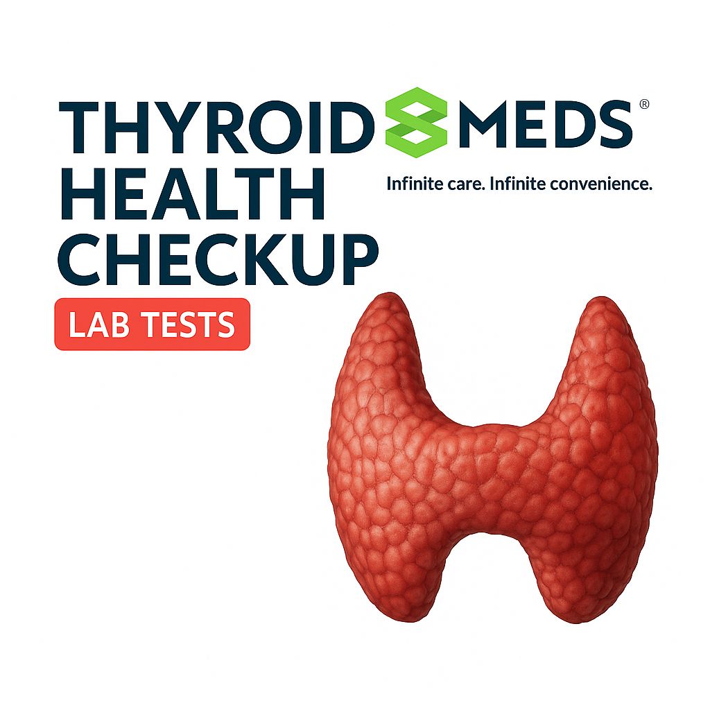
🧪 Thyroxine-Binding Globulin (TBG) Test The Thyroxine-Binding Globulin (TBG) test measures the amount of TBG protein in your blood. TBG is a transport protein that binds to thyroid hormones (T3 and T4) and carries them through the bloodstream. It’s not a routine thyroid function test, but it’s useful in special cases when thyroid hormone levels seem abnormal but TSH, Free T3, and Free T4 don’t fully explain symptoms. 🧬 What Is TBG? Feature Description Full Name Thyroxine-Binding Globulin Main Role Binds to and transports T3 and T4 Bound Hormones Are inactive (only free hormones are active) Produced by Liver 📊 Normal Reference Range (May vary by lab) Test Normal Range TBG 15 – 30 µg/mL (or) 1.3 – 2.0 mg/L 📈 High or 📉 Low TBG – What It Means 📈 Increased TBG More hormone is bound, less Free T4/T3 available (but Total T4/T3 may be high) Causes: Pregnancy (due to estrogen) Estrogen therapy (e.g., oral contraceptives) Hepatitis Genetic increase in TBG 📉 Decreased TBG Less hormone is bound, more Free T4/T3 relative to Total Causes: Liver disease Nephrotic syndrome Corticosteroid use Androgens or anabolic steroids Genetic TBG deficiency 🧪 Why Is the TBG Test Done? Use Case Purpose Unexplained high or low Total T4/T3 To assess if altered due to binding issues Pregnancy or hormone therapy Understand changes in thyroid tests Suspected inherited TBG disorder Evaluate genetic binding protein conditions 🧾 Example Lab Report Test Result Reference Range Thyroxine-Binding Globulin 28 µg/mL 15 – 30 µg/mL ✅ Interpretation: TBG within normal limits — does not affect total/free T3/T4 interpretation. 🧠 Important Notes TBG does not measure thyroid hormone levels directly. Use Free T3 and Free T4 to assess actual thyroid hormone activity. If Total T4 is high and Free T4 is normal, elevated TBG might be the reason. ✅ Summary Feature Details Test Thyroxine-Binding Globulin (TBG) Function Binds and transports thyroid hormones High TBG Seen in pregnancy, estrogen therapy Low TBG Seen in liver disease, steroid use Best Used With Total and Free T4/T3 levels
₹6000
TVS SCAN ( Transvaginal Sonography )
Report in 1 HOUR
🧪 TVS Scan (Transvaginal Sonography) Transvaginal Sonography (TVS) is a pelvic ultrasound scan performed using a probe inserted into the vagina. It provides high-resolution images of female reproductive organs, including the uterus, endometrium, ovaries, fallopian tubes, and cervix. 🔬 Test Overview Parameter Details Test Name Transvaginal Sonography (TVS) Also Known As Endovaginal Ultrasound Sample Type Imaging (vaginal ultrasound probe) Fasting Required? ❌ Not required Bladder Status ❌ Empty bladder preferred Duration 10–20 minutes Performed By Radiologist or OB/GYN with ultrasonography training Radiation? ❌ None 🧬 Clinical Uses of TVS Evaluate abnormal uterine bleeding or irregular cycles Monitor follicular development in infertility treatments Detect ovarian cysts, fibroids, polyps, or ectopic pregnancy Assess endometrial thickness (e.g., in postmenopausal bleeding) Confirm early pregnancy and fetal heartbeat Diagnose pelvic inflammatory disease (PID) Assist in IUD placement evaluation 📊 Common Parameters Assessed Structure What’s Measured/Observed Uterus Size, shape, fibroids, position Endometrium Thickness, uniformity (varies with menstrual phase) Ovaries Follicle count, cysts, masses Pouch of Douglas Presence of fluid (may suggest infection or rupture) Cervix Length, funneling (in pregnancy), abnormalities 📅 When Is It Performed? Day 2–5 of cycle: For baseline fertility assessment Mid-cycle: To monitor ovulation or follicular growth During pregnancy: For early viability scan (around 6–9 weeks) Postmenopausal: To evaluate endometrial thickness or bleeding ✅ Summary Test Transvaginal Sonography (TVS) Purpose Detailed pelvic organ imaging Safe for Pregnancy ✅ Yes – especially early pregnancy Invasive? Minimally (vaginal probe insertion) Radiation ❌ None
₹1200
🧪 Food Allergy Profile (Vegetarians) The Food Allergy Profile (Vegetarian) is a blood test that screens for IgE-mediated allergic reactions to common vegetarian foods. It helps identify which plant-based foods may be triggering allergic symptoms such as: Skin rashes (eczema, hives) Gastrointestinal upset (bloating, diarrhea) Respiratory issues (sneezing, wheezing) Anaphylaxis (in rare severe cases) 🔬 Test Overview Parameter Details Test Name Vegetarian Food Allergy Panel (IgE) Sample Type Blood (Serum) Method ImmunoCAP / ELISA (specific IgE antibodies) Fasting Required? ❌ Not required Turnaround Time 3–5 working days Units kU/L (kilo units per liter) 🍅 Typical Vegetarian Food Allergens Included Depending on the lab, panels may vary slightly, but commonly include: Food Item Examples Legumes Soybean, Lentil, Pea, Chickpea Grains Wheat, Rye, Barley, Oats Nuts & Seeds Peanut, Almond, Walnut, Sesame Vegetables Tomato, Potato, Spinach, Eggplant Fruits Banana, Apple, Kiwi, Mango Others Mustard, Coconut, Corn, Garlic 📊 Interpretation of Results (IgE Levels) IgE Level (kU/L) Class Allergy Risk < 0.35 Class 0 Negative 0.35 – 0.69 Class 1 Low/Borderline 0.70 – 3.49 Class 2 Moderate sensitivity 3.50 – 17.49 Class 3 High sensitivity ≥17.5 Class 4+ Very high sensitivity ⚠️ Note: A positive IgE test doesn't always mean clinical allergy — correlation with symptoms and physician evaluation is essential. 🧬 When Is This Test Recommended? Unexplained allergic symptoms after vegetarian meals History of eczema, asthma, or hay fever Evaluation for oral allergy syndrome Diet planning for suspected food allergies Pediatric allergy workup for plant-based foods ✅ Summary Test Vegetarian Food Allergy Panel (IgE) Sample Serum (Blood) Analyzes Specific IgE to plant-based foods Useful For Identifying vegetarian food allergens Next Steps Diet modification, allergist referral, oral food challenge
₹3500
₹4500
A hemoglobin test is a common blood test that measures the amount of hemoglobin in your blood. Hemoglobin is a protein in red blood cells responsible for carrying oxygen from your lungs to the rest of your body and returning carbon dioxide back to the lungs. 🔬 Why It's Done: Doctors order this test to: Check for anemia (low hemoglobin) Monitor a known blood disorder (e.g. sickle cell, thalassemia) Assess general health as part of a complete blood count (CBC) Check for polycythemia (high hemoglobin) 📉 Normal Hemoglobin Ranges: These values can vary slightly between labs: Group Normal Range (g/dL) Men 13.8 – 17.2 Women 12.1 – 15.1 Children 11.0 – 16.0 Pregnant women 11.0 – 12.0 (lower) 🔻 Low Hemoglobin (Anemia) Causes: Iron deficiency Vitamin B12 or folate deficiency Chronic diseases (kidney, cancer) Blood loss (injury, menstruation) Bone marrow problems 🔺 High Hemoglobin Causes: Dehydration Lung or heart disease Living at high altitudes Smoking Polycythemia vera (a bone marrow disorder) 🧪 How the Test Is Done: A healthcare provider draws a blood sample from your arm. No fasting or special preparation is usually needed (unless part of a broader test panel).
₹1
FOOD ALLERGY PROFILE ( FOR NON VEGETARIANS )
Report in 48 HOURS
🧪 Food Allergy Profile (Non-Vegetarian) The Food Allergy Profile for Non-Vegetarians is a diagnostic blood test that detects IgE antibodies against common non-vegetarian food allergens. It helps identify food items that may trigger allergic reactions like skin rashes, gastrointestinal discomfort, respiratory issues, or anaphylaxis. 🔬 Test Overview Parameter Details Test Name Food Allergy Profile – Non-Vegetarian Sample Type Blood (serum) Method Immunoassay (e.g. ELISA, CLIA, ImmunoCAP) Fasting Required? ❌ Not required Turnaround Time 3–7 days Analytes Total IgE + Specific IgE to non-vegetarian foods 🍗 Non-Vegetarian Allergens Commonly Included Category Examples Tested Meat Chicken, Mutton, Beef, Pork Eggs Egg White, Egg Yolk Seafood Fish (Tuna, Salmon, Cod, Sardine), Shrimp, Crab, Lobster, Squid Dairy Proteins Cow Milk, Buffalo Milk, Casein, Whey (often included if requested) 📊 Result Interpretation (Specific IgE Levels) IgE Level (kU/L) Class Interpretation < 0.35 Class 0 Negative 0.35–0.70 Class 1 Low sensitivity (possible sensitization) 0.71–3.50 Class 2 Moderate allergy likelihood 3.51–17.50 Class 3 High sensitivity >17.50 Class 4–6 Strong to severe sensitivity ⚠️ Possible Symptoms of Non-Veg Food Allergy Itchy skin, hives, eczema Nausea, vomiting, bloating Wheezing, nasal congestion Swelling of lips or throat Anaphylaxis (in severe seafood/meat allergy cases) 🧬 When to Consider This Test History of reactions after eating meat, seafood, or eggs Unexplained skin or digestive issues Family history of allergies Atopic conditions like asthma, eczema, or allergic rhinitis ✅ Summary Test Food Allergy Profile (Non-Vegetarian) Purpose Detect IgE-mediated allergies to non-veg foods Includes Meat, Eggs, Seafood (customizable) Method IgE Immunoassay (e.g., ELISA, CLIA) Fasting Needed No Result Format Quantitative IgE levels with Class grading Next Steps Allergen elimination, antihistamines, epinephrine (if severe)
₹4500
FOOD TOLERANCE TEST
Report in 5 DAYS
🧪 Food Tolerance Test – Overview & Clinical Insight A Food Tolerance Test assesses how your body reacts to specific foods or food components—not through an immune-mediated allergic reaction, but via non-immunologic responses, commonly referred to as food sensitivities or intolerances. These reactions are typically delayed and can affect digestion, metabolism, or other physiological systems. 🔬 Types of Food Tolerance Tests Test Type What It Measures Common Uses Lactose Tolerance Test Hydrogen in breath or blood glucose after lactose ingestion Diagnose lactose intolerance Fructose Tolerance Test Hydrogen breath test or symptom tracking Diagnose fructose malabsorption Gluten Sensitivity Testing Excludes celiac markers (tTG, EMA) + symptom tracking Diagnose non-celiac gluten sensitivity Food-Specific IgG Testing IgG antibodies to 90–270 foods (controversial) Evaluate delayed food sensitivities (caution) Elimination Diet & Challenge Gold standard: Remove suspected foods, reintroduce systematically Identify problematic foods via symptoms Histamine Intolerance Test DAO enzyme activity + histamine levels Diagnose histamine intolerance (e.g., red wine, cheese) FODMAP Sensitivity Test Often done via elimination & breath testing Identify fermentable carb intolerance ⚠️ Important Note on IgG Food Testing Many commercial "Food Intolerance Tests" check IgG antibodies to various foods. Medical consensus (e.g., AAAAI, EAACI): IgG levels often reflect exposure, not intolerance. Elevated IgG does not prove intolerance. These tests may lead to unnecessary food restrictions if misinterpreted. 📊 Typical Food Intolerance Symptoms System Symptoms GI Tract Bloating, diarrhea, constipation, gas, nausea Neurological Headache, migraine, foggy thinking, fatigue Skin Eczema, rashes, acne, itching Respiratory Nasal congestion, sinusitis Mood/Behavioral Irritability, depression, anxiety 🧬 Common Intolerances to Test/Monitor Food Component Example Sources Test Type Lactose Milk, cheese, ice cream Lactose breath test Gluten (non-celiac) Bread, pasta, wheat Exclusion & challenge Fructose Fruits, honey, soft drinks Fructose breath test Histamine Aged cheese, wine, smoked meat DAO enzyme test Salicylates Spices, tomatoes, berries Elimination diet FODMAPs Onion, garlic, beans, apples FODMAP diet testing Additives (sulfites, MSG) Packaged foods, wine, sauces Symptom challenge testing ✅ Best Practices Gold Standard: Elimination and food challenge under dietitian or doctor supervision. IgG Food Panel: May provide clues but should not be used alone for diagnosis. Combine: Symptom diary + targeted tests (like lactose breath test) for accuracy. ✅ Summary Table Test Food Tolerance Test Used For Identifying food sensitivities and intolerances Types IgG panels, hydrogen breath tests, elimination diets Best For Digestive complaints, skin issues, fatigue, migraines Caution IgG levels ≠ clinical intolerance (not allergy-specific) Clinical Guidance Always interpret with physician/dietitian support
₹15898
FOOD ALLERGY - MILK PANEL ( IgE TEST )
Report in 6 DAYS
🧪 Food Allergy – Milk Panel (IgE Test) The Milk Allergy Panel detects IgE antibodies specific to various milk proteins. It helps diagnose IgE-mediated allergic reactions to cow’s milk and related dairy products, which are common in infants, children, and some adults. 🔬 Test Overview Parameter Details Test Name Milk Allergy Panel (IgE) Sample Type Blood (Serum) Method Immunoassay (e.g., ImmunoCAP, ELISA) Fasting Required? ❌ Not required Turnaround Time 2–4 working days Units kU/L (kilo units per liter) 🥛 Milk Proteins Commonly Tested Allergen Component Description Whole Cow’s Milk General milk protein profile Casein Heat-stable protein, major cause of persistent allergy α-Lactalbumin Whey protein, often triggers symptoms β-Lactoglobulin Whey protein, common allergen in infants Bovine Serum Albumin (BSA) Minor component; cross-reactive with beef 📊 Result Interpretation (IgE Levels) IgE Level (kU/L) Class Risk of Allergy < 0.35 Class 0 Negative 0.35 – 0.69 Class 1 Borderline/Low 0.70 – 3.49 Class 2 Moderate 3.50 – 17.49 Class 3 High 17.50 – 49.99 Class 4 Very High ≥ 50 Class 5–6 Extremely High 🔎 Note: IgE presence indicates sensitization, but clinical correlation is essential—not all positive tests indicate true allergy. 🧬 Common Symptoms of Milk Allergy Skin: Eczema, hives, itching GI: Vomiting, colic, diarrhea, bloating Respiratory: Wheezing, nasal congestion Severe: Anaphylaxis (rare but serious) ❗ Differentiating Milk Allergy vs. Lactose Intolerance Milk Allergy Lactose Intolerance Immune system (IgE-mediated) Enzyme deficiency (lactase) Involves proteins (casein, whey) Involves sugar (lactose) Onset: Minutes to hours Onset: Hours after ingestion Symptoms: Rash, swelling, wheeze, GI Symptoms: Bloating, gas, diarrhea ✅ Summary Test Milk Allergy Panel (IgE-based) Components Analyzed Whole milk, casein, α-lactalbumin, β-lactoglobulin, BSA Purpose Diagnose IgE-mediated milk protein allergy Population Commonly used in infants, children, allergic individuals Follow-Up Elimination diet, allergist consultation, possible oral food challenge
₹1380
ALLERGY DUST PANEL ( Serum Specific IgE Test )
Report in 6 DAYS
🧪 Allergy Dust Panel (Serum Specific IgE Test) The Dust Allergy Panel is a specific IgE test that identifies allergic sensitization to common indoor allergens, especially house dust mites, which are among the leading causes of allergic rhinitis, asthma, and eczema. 🔬 Test Overview Parameter Details Test Name Dust Allergy Panel (Specific IgE) Sample Type Blood (serum) Method Immunoassay (e.g., ImmunoCAP, ELISA) Fasting Required? ❌ Not required Turnaround Time 3–5 working days Units kU/L (kilo units per liter of specific IgE) 🧫 Common Allergens in Dust Panel Allergen Source Dermatophagoides pteronyssinus House dust mite (European) Dermatophagoides farinae House dust mite (American) Blomia tropicalis Tropical dust mite Cockroach Antigen (e.g., Bla g 1) Kitchen/bathroom surfaces Storage Mite Mix (e.g., Tyrophagus, Acarus) Stored food/grains House Dust (Mixed Sample) Composite of dust particles from home Mold Spores (Optional) May be added depending on lab (e.g., Alternaria) 📊 IgE Class Reference Range IgE Level (kU/L) Class Clinical Interpretation < 0.35 Class 0 Negative 0.35 – 0.69 Class 1 Low Sensitization 0.70 – 3.49 Class 2 Moderate 3.50 – 17.49 Class 3 High 17.50 – 49.99 Class 4 Very High ≥ 50.0 Class 5–6 Extremely High ⚠️ A positive result suggests sensitization, but clinical correlation is essential to determine if it's causing symptoms. 🤧 Common Symptoms of Dust Allergy Sneezing, runny or blocked nose (rhinitis) Itchy, watery eyes Chronic cough Asthma (wheezing, shortness of breath) Eczema flares Sinus pressure or headaches 🧬 Clinical Utility Confirm house dust mite or cockroach allergy Aid in diagnosing allergic asthma or rhinitis Guide treatment including allergen avoidance and immunotherapy Evaluate indoor air quality risk 🧹 Dust Allergy Management Tips Use HEPA filters and air purifiers Encasement covers for mattresses and pillows Remove carpets, stuffed toys, and other dust collectors Frequent cleaning with damp cloth and hot water washing Control indoor humidity (<50%) ✅ Summary Panel Name Dust Allergy Panel (Specific IgE) Purpose Detect sensitization to indoor dust allergens Sample Blood (serum) Includes House dust mites, cockroach, storage mites Recommended For Allergic rhinitis, asthma, eczema sufferers
₹3898
ALLERGY ANIMAL PANEL ( Serum Specific IgE Test )
Report in 6 DAYS
🧪 Allergy Animal Panel (Serum Specific IgE Test) The Animal Allergy Panel detects IgE antibodies in blood against common animal allergens, helping to diagnose IgE-mediated allergic reactions to animal dander, saliva, and urine. This test is commonly used for individuals with respiratory symptoms (e.g., asthma, rhinitis) that worsen around animals. 🔬 Test Overview Parameter Details Test Name Animal Allergy Panel (Specific IgE) Sample Type Blood (serum) Method Immunoassay (e.g., ImmunoCAP, ELISA) Fasting Required? ❌ Not required Turnaround Time 3–5 working days Units kU/L (kilo units per liter of IgE) 🐾 Common Allergens Included Allergen Source Cat Dander (Fel d 1) Skin, saliva, fur (cats) Dog Dander (Can f 1, Can f 5) Saliva, skin (dogs) Horse Dander Hair, sweat, skin Mouse Urine Protein Bedding, air in infested spaces Rat Epithelium/Urine Lab exposure, pet rats Guinea Pig Epithelium Fur, skin flakes Rabbit Epithelium Skin, fur Feather Mix (e.g., Chicken, Duck, Goose) Bedding, pillows Sheep Wool Clothing, blankets 📊 IgE Class Reference Range IgE Level (kU/L) Class Clinical Significance < 0.35 Class 0 Negative 0.35 – 0.69 Class 1 Low Sensitization 0.70 – 3.49 Class 2 Moderate 3.50 – 17.49 Class 3 High 17.50 – 49.99 Class 4 Very High ≥ 50 Class 5–6 Extremely High ⚠️ Note: Positive results indicate sensitization but may not always cause symptoms. Correlate clinically. 🤧 Common Symptoms of Animal Allergy Sneezing, nasal congestion Itchy, watery eyes Cough, wheezing, asthma Skin rashes, hives Exacerbation of eczema 🧬 Clinical Utility Diagnose pet or occupational animal allergies Guide allergen avoidance strategies Support immunotherapy decisions Evaluate risk of asthma or allergic rhinitis ✅ Summary Panel Name Animal Allergy Panel (IgE) Components Dander from cats, dogs, rodents, horses, birds, etc. Test Purpose Detect allergic sensitization to animals Population Individuals with respiratory or skin symptoms triggered by animal exposure Follow-Up Allergen avoidance, antihistamines, or allergy shots (immunotherapy)
₹3500
ASPERGILLUS FUMIGATUS TEST
Report in 48 HOURS
🧪 Aspergillus fumigatus Test Aspergillus fumigatus is a species of fungus that can cause a range of illnesses, especially in people with weakened immune systems, lung disease, or allergies. Laboratory testing for Aspergillus fumigatus is used to detect infection, colonization, or allergic responses. 🧬 What Is Aspergillus fumigatus? A common mold found in the environment (soil, decaying leaves, compost). Normally harmless, but can cause illness in immunocompromised individuals or those with lung conditions. 🧪 Common Tests for Aspergillus fumigatus Test Name Purpose Aspergillus IgG antibody Detects chronic or subacute infection (e.g., CPA) Aspergillus IgE antibody Detects allergic reaction (e.g., ABPA) Aspergillus Galactomannan Detects fungal antigen in blood or BAL fluid; helpful in invasive aspergillosis Fungal culture Grows the fungus from sputum or tissue PCR test Detects Aspergillus DNA CT scan (not a lab test) Helps identify lung damage from fungus 📊 Interpretation of Results Condition Typical Lab Findings Allergic Bronchopulmonary Aspergillosis (ABPA) ↑ IgE, ↑ Aspergillus-specific IgE/IgG Chronic Pulmonary Aspergillosis (CPA) ↑ Aspergillus IgG, positive culture, lung cavity on CT Invasive Aspergillosis + Galactomannan, PCR, possibly culture; often in ICU or neutropenic patients Aspergilloma (fungus ball) CT scan shows a mass; IgG may be elevated Colonization Positive culture but no signs of active infection 📈 Sample Reference Ranges Test Normal Range Aspergillus IgG < 10–15 mg/L (varies by lab) Aspergillus IgE < 0.35 kU/L (cutoff varies) Galactomannan (serum) < 0.5 index value Positive IgG or IgE results suggest immune sensitization or infection, depending on clinical context. ⚠️ Who Should Be Tested? Patients with: Persistent cough or hemoptysis Asthma with worsening symptoms Bronchiectasis Lung cavities on imaging Weakened immunity (chemotherapy, transplant, HIV/AIDS) Non-resolving pneumonia ✅ Summary Feature Details Organism Aspergillus fumigatus Diseases ABPA, CPA, invasive aspergillosis, aspergilloma Tests IgG, IgE, Galactomannan, PCR, Culture Indications Persistent lung symptoms, immunocompromised status
₹1000
AFB TEST ( ACID-FAST BACILLUS )
Report in 24 Hours
🧪 AFB Test (Acid-Fast Bacillus Test) The AFB test is used to detect Mycobacterium species, especially Mycobacterium tuberculosis (TB) — the bacteria that causes tuberculosis. "Acid-fast bacilli" refers to the unique staining characteristics of these bacteria. ✅ Why Is the AFB Test Done? To diagnose active tuberculosis (TB) or monitor treatment response in patients with: Persistent cough lasting more than 2 weeks Weight loss Fever, night sweats Coughing up blood (hemoptysis) Suspicion of pulmonary or extrapulmonary TB 🔬 Types of AFB Tests Type Sample Purpose AFB Smear (Microscopy) Sputum, pus, body fluid Quick, initial detection AFB Culture Sputum, blood, tissue, fluids Confirms TB and identifies drug resistance (takes weeks) AFB PCR / NAAT Sputum, blood, CSF Rapid genetic test to confirm TB and resistance 📊 Interpretation of Results Result Meaning Positive AFB smear Likely TB infection (or another Mycobacterium); needs culture confirmation Negative smear TB not ruled out — repeat testing or use PCR/culture Positive culture Definitive TB diagnosis Positive PCR (NAAT) Confirms TB DNA; fast and specific 🧪 Sample Requirements Sputum (most common): Early morning, deep cough, 3 consecutive days Other samples: Urine, CSF, lymph node aspirate, gastric lavage, etc. (for extrapulmonary TB) 🧍♂️🧍♀️ Who Should Get an AFB Test? Anyone with suspected TB symptoms Close contacts of TB patients People with HIV, diabetes, or weakened immunity Healthcare workers or people in high-risk environments 🔁 Often Ordered With: Test Use Chest X-ray To look for TB lesions Tuberculin Skin Test (TST) or IGRA TB exposure screening CBNAAT (GeneXpert) Rapid molecular TB test + rifampicin resistance HIV test TB often co-occurs with HIV ESR, CBC Supportive information on inflammation or infection 📌 Summary Test Detects Use AFB Smear Acid-fast bacilli under microscope Initial screen AFB Culture Grows the organism Gold standard AFB PCR / NAAT DNA of TB bacteria Fast, confirmatory test
₹900
MANTOUX TEST ( Tuberculin Skin Test – TST )
Report in 24 HOURS
: 🧪 Mantoux Test (Tuberculin Skin Test – TST) The Mantoux test is a diagnostic tool used to detect latent or active tuberculosis (TB) infection. It involves intradermal injection of Purified Protein Derivative (PPD) and checking the body’s immune response. 🔬 Test Overview Parameter Details Test Name Mantoux Test / Tuberculin Skin Test (TST) Method Intradermal injection of 0.1 mL PPD (5 TU) Injection Site Inner forearm (volar surface) Sample Type No blood sample; intradermal reaction Fasting Required? ❌ Not required Result Time 48 to 72 hours after injection 📊 Result Interpretation After 48–72 hours, the induration (raised, hardened area, not redness) is measured in millimeters: Induration Size Interpretation ✅ ≥5 mm Positive in high-risk individuals (HIV+, recent TB contact, immunosuppressed) ⚠️ ≥10 mm Positive in moderate-risk groups (healthcare workers, recent immigrants, children <5) ❗ ≥15 mm Positive in healthy individuals with no known risk factors 🔻 <5 mm Usually negative, but may require repeat testing in high-risk or immunocompromised cases ⚠️ Limitations False Positives: Prior BCG vaccination Exposure to non-tuberculous mycobacteria False Negatives: Weakened immunity (HIV, steroids, cancer) Very recent TB infection Very young or elderly individuals 🧬 Alternative: IGRA Blood Test The Interferon Gamma Release Assay (IGRA) is a blood test alternative (e.g., QuantiFERON-TB Gold) that: Is not affected by BCG vaccine Requires only one visit Useful in BCG-vaccinated individuals ✅ Summary Test Mantoux Test (TST) Purpose Detect latent or active TB infection Type Skin test with PPD injection Read After 48–72 hours Positive If Induration ≥5–15 mm (based on risk group) Follow-Up Chest X-ray, sputum test, IGRA if needed
₹200
FOOD ALLERGY PROFILE ( VEGETARIANS & NON VEGETARIANS)
Report in 48 HOURS
🧪 Food Allergy Profile (Vegetarians & Non-Vegetarians) The Food Allergy Profile (Vegetarian + Non-Vegetarian) is a comprehensive blood test that detects IgE-mediated allergic responses to a wide variety of common foods across both vegetarian and non-vegetarian categories. It is useful in diagnosing food allergies that can cause skin rashes, digestive issues, respiratory problems, or even anaphylaxis. 🔬 Test Overview Parameter Details Test Name Food Allergy Profile – Vegetarian & Non-Vegetarian Sample Type Blood (serum) Method Immunoassay (e.g. ELISA, ImmunoCAP, CLIA) Fasting Required? ❌ Not necessary Turnaround Time 3–7 days (depending on lab and panel size) Analytes Total IgE + Specific IgE for multiple food allergens 🥦 Common Vegetarian Food Allergens Tested Cereals/Grains: Wheat, Rice, Oats, Corn, Barley Pulses: Soybean, Lentils, Chickpeas, Peanuts Vegetables: Tomato, Potato, Brinjal, Spinach Fruits: Banana, Apple, Mango, Strawberry, Citrus fruits Nuts/Seeds: Almonds, Cashew, Walnut, Sesame, Mustard Spices: Turmeric, Cumin, Clove 🍗 Common Non-Vegetarian Food Allergens Tested Meat: Chicken, Mutton, Beef, Pork Seafood: Prawns, Crab, Lobster, Salmon, Tuna, Sardines Eggs: Egg white, Egg yolk Dairy: Cow milk, Buffalo milk, Casein, Whey 📊 Interpretation of Results IgE Level (kU/L) Class Interpretation < 0.35 Class 0 Negative 0.35–0.70 Class 1 Low sensitivity 0.71–3.50 Class 2 Moderate sensitivity 3.51–17.50 Class 3 High sensitivity > 17.50 Class 4–6 Very high sensitivity (likely clinical) ⚠️ Symptoms of Food Allergies Itching or swelling of mouth/throat Skin rashes, hives, or eczema Nausea, vomiting, diarrhea Wheezing, nasal congestion Anaphylaxis (life-threatening in severe cases) 🧬 Why This Test Is Important Diagnoses hidden causes of chronic allergy symptoms Helps plan an elimination diet Useful in children with eczema, asthma, or food aversions Identifies allergens that may worsen autoimmune or GI conditions ✅ Summary Test Food Allergy Profile (Veg + Non-Veg) Method IgE immunoassay No. of Allergens Varies (typically 30–100+) Best For Suspected food-triggered allergies or intolerance Preparation No fasting required Result Format Specific IgE values + Class rating
₹6000
HUMAN GROWTH HORMONE ( HGH ) TEST
Report in 24 HOURS
🧪 Human Growth Hormone (HGH) Test The Human Growth Hormone (HGH) test measures the amount of growth hormone in your blood. Growth hormone is produced by the pituitary gland and plays a critical role in growth, metabolism, muscle and bone development, and tissue repair. 🔬 Test Overview Parameter Details Test Name Human Growth Hormone (HGH), Serum Sample Type Blood (Serum) Method Chemiluminescent Immunoassay (CLIA) / ELISA Fasting Required? ✅ Yes (usually 8–12 hours) Timing Often measured in the morning or during stimulation/suppression tests Turnaround Time 1–3 days Units ng/mL (nanograms per milliliter) 📊 Normal Reference Ranges Group HGH Normal Range (may vary slightly by lab) Men <5 ng/mL Women <10 ng/mL Children 10–50 ng/mL (can be higher during growth spurts) 🧠 Note: HGH levels fluctuate throughout the day, so random measurements can be misleading. Specialized tests (see below) are more reliable for diagnosis. 🧪 Associated Tests (for accurate diagnosis) Test Purpose GH stimulation test Diagnose GH deficiency (e.g., using insulin, arginine) GH suppression test Diagnose GH excess (e.g., acromegaly, gigantism) IGF-1 test (Insulin-like Growth Factor 1) More stable marker reflecting average HGH activity ⚠️ Clinical Uses of HGH Testing ⬇️ Low HGH Levels (Deficiency): Short stature in children Delayed puberty Growth hormone deficiency in adults (fatigue, decreased muscle mass) Pituitary disorders (e.g., tumors, trauma) ⬆️ High HGH Levels (Excess): Acromegaly (in adults): enlarged hands, feet, facial bones Gigantism (in children): abnormal height and growth Pituitary adenomas (GH-secreting tumors) ✅ Summary Test Human Growth Hormone (HGH) – Serum Purpose Diagnose GH deficiency or excess Key Markers HGH + IGF-1 + Stimulation/Suppression tests Sample Type Blood (serum) – often fasting Next Steps If abnormal, do dynamic testing (stim/supp)
₹900
PLATELET COUNT TEST ( Thrombocyte Count )
Report in 24 HOURS
🩸 Platelet Count Test (Thrombocyte Count) ✅ What is a Platelet Count Test? A platelet count test measures the number of platelets (thrombocytes) in your blood. Platelets are small cell fragments that help with blood clotting and prevent excessive bleeding when injuries occur. 🔬 Normal Platelet Count Range: Age Group Normal Range Adults 150,000 – 450,000 /µL (or 150–450 × 10⁹/L) Children Slightly higher variation possible Pregnancy May be slightly lower, especially in 3rd trimester 📈 High Platelet Count (Thrombocytosis): Platelet count > 450,000 /µL Possible Causes: Infections Inflammation (e.g., rheumatoid arthritis, IBD) Iron deficiency anemia After surgery or trauma Chronic myeloproliferative disorders (e.g., essential thrombocythemia) Cancer Symptoms (if any): Often none, but sometimes: Headaches Dizziness Blood clots (rare) 📉 Low Platelet Count (Thrombocytopenia): Platelet count < 150,000 /µL Possible Causes: Dengue fever, viral infections (e.g., HIV, Hepatitis) Bone marrow disorders Autoimmune diseases (e.g., ITP, lupus) Medications (e.g., chemotherapy, antibiotics, heparin) Alcoholism Sepsis Pregnancy-related conditions Symptoms: Easy bruising Prolonged bleeding Petechiae (small red/purple spots under skin) Gum or nose bleeding 🧪 Why is the Test Done? To evaluate: Unexplained bruising or bleeding Blood disorders (like dengue, leukemia, aplastic anemia) Bone marrow health Response to treatments (e.g., chemotherapy, platelet transfusion) As part of a Complete Blood Count (CBC) 🧍♂️🧍♀️ Test Details: Sample: Blood (venipuncture) Fasting: Not required Results: Usually available within a few hours 📝 Summary: Platelet Count (/µL) Interpretation < 150,000 Low (Thrombocytopenia) – Risk of bleeding 150,000–450,000 Normal > 450,000 High (Thrombocytosis) – Risk of clotting
₹150
HELICOBACTER PYLORI ( H.PYLORI ) ANTIGEN TEST - STOOL
Report in 2 DAYS
🧪 Helicobacter pylori (H. pylori) Antigen – Stool Test The H. pylori stool antigen test detects the presence of H. pylori bacteria in a stool sample. It is a non-invasive, highly accurate test used for initial diagnosis and post-treatment confirmation of H. pylori infection — a common cause of gastritis, peptic ulcers, and sometimes stomach cancer. 🧬 What is H. pylori? A spiral-shaped bacterium that infects the stomach lining. Often acquired in childhood. Can cause: Gastritis Peptic ulcers Gastric cancer (rare, long-term complication) Dyspepsia (indigestion) 📊 Reference Range Test Result Interpretation Negative No H. pylori antigen detected (normal) Positive H. pylori infection present Equivocal Borderline result; may require repeat test 🧾 Example Lab Report Test Result Reference Range H. pylori Stool Antigen Positive Negative Interpretation Infection present – needs treatment 🧪 Test Use Cases Purpose Description Diagnosis Detect active H. pylori infection Post-treatment testing Confirm eradication 4+ weeks after therapy Screening In patients with ulcer symptoms or family history 📋 Before the Test To avoid false-negative results, follow these guidelines: Medication Stop Before Test Antibiotics ≥ 4 weeks PPIs (e.g., omeprazole) ≥ 2 weeks Bismuth compounds ≥ 4 weeks 💊 If Positive – Treatment (Triple Therapy) A typical 14-day regimen may include: PPI (e.g., omeprazole or pantoprazole) Amoxicillin Clarithromycin Alternatives exist if resistant strains or allergies are present. ✅ Summary Feature Detail Test H. pylori stool antigen Sample Stool Result: Positive Active infection – treat accordingly Result: Negative No current infection Best for Initial diagnosis and post-treatment follow-up Avoid false results Discontinue PPIs & antibiotics beforehand
₹3000
VITAMIN B COMPLEX PROFILE TEST
Report in 1 WEEK
💊 Vitamin B Complex Profile Test The Vitamin B Complex Profile measures the levels of multiple B vitamins in your body — a group of essential water-soluble vitamins that play key roles in energy metabolism, nervous system function, red blood cell production, and DNA synthesis. 🧪 What’s Included in a Typical B Complex Profile? Vitamin Also Known As Key Function B1 Thiamine Nerve function, carbohydrate metabolism B2 Riboflavin Energy production, skin and eye health B3 Niacin Energy metabolism, DNA repair B5 Pantothenic Acid Fatty acid metabolism, hormone synthesis B6 Pyridoxal Phosphate Neurotransmitter synthesis, amino acid metabolism B7 Biotin Hair, skin, nails; carb/fat/protein metabolism B9 Folate (Folic Acid) DNA synthesis, pregnancy support, red blood cell formation B12 Cobalamin Nerve function, red blood cells, DNA synthesis Some labs may test only key vitamins like B1, B2, B6, B9, and B12 unless specifically requested. 📊 Reference Ranges (May Vary by Lab) Vitamin Normal Range (Common Units) B1 (Thiamine) 70 – 180 nmol/L (whole blood) B2 (Riboflavin) 5 – 50 µg/L (plasma) B3 (Niacin) 0.5 – 8.5 µg/mL (blood or urine) B5 (Pantothenic Acid) 1.6 – 2.7 µmol/L B6 (Pyridoxal Phosphate) 5 – 30 µg/L B7 (Biotin) 200 – 1200 ng/L B9 (Folate) > 4 ng/mL (serum) or > 140 ng/mL (RBC) B12 (Cobalamin) 200 – 900 pg/mL 🔍 Why This Test Is Ordered Reason Explanation 🧠 Neuropathy, fatigue, confusion Suspect B1, B6, or B12 deficiency 👩🍼 Pregnancy monitoring Folate (B9) and B12 are critical for fetal health 🍺 Alcohol use disorder Risk of B vitamin deficiencies 🥗 Vegan/vegetarian diet Risk of low B12 intake 💉 Malabsorption disorders Crohn’s, celiac, bariatric surgery 💊 Medication effects Metformin, PPIs, isoniazid may deplete B vitamins ⚠️ Symptoms of B Vitamin Deficiency B Vitamin Deficiency Symptoms B1 Fatigue, nerve pain, confusion, Wernicke-Korsakoff syndrome B2 Cracks at mouth corners, sore throat, red eyes B3 Pellagra (dermatitis, diarrhea, dementia) B5 Fatigue, mood disturbances (rare) B6 Irritability, depression, neuropathy B7 Hair thinning, brittle nails, skin rash B9 Anemia, birth defects, poor concentration B12 Anemia, memory loss, numbness/tingling, glossitis 🩺 How Is the Test Done? Parameter Details Sample Blood (serum/plasma or whole blood) Fasting? Yes, fasting preferred (8–10 hours) Processing Time 1–5 days depending on lab 🧾 Sample Test Report (Summary) Vitamin Result Reference Range Status B1 60 nmol/L 70–180 nmol/L Deficient B2 12 µg/L 5–50 µg/L Normal B6 8.3 µg/L 5–30 µg/L Normal B9 3.2 ng/mL >4 ng/mL Low B12 154 pg/mL 200–900 pg/mL Low 🟥 Interpretation: B1, B9, and B12 deficiencies — commonly seen in alcohol-related malnutrition or vegetarian diets. ✅ Summary Feature Detail Test Name Vitamin B Complex Profile Includes B1 through B12 (most or all B vitamins) Use Diagnose deficiency or toxicity Fasting Needed Preferred Toxicity Risk Low (excess excreted in urine, except B6 in very high doses)
₹8000
VITAMIN B2 TEST ( RIBOFLAVIN )
Report in 10 DAYS
💊 Vitamin B2 (Riboflavin) Test The Vitamin B2 (Riboflavin) Test measures levels of riboflavin in your body — a water-soluble B vitamin essential for energy production, cellular metabolism, red blood cell function, and maintaining healthy skin, eyes, and nerves. 🧬 Why Is This Test Done? Clinical Use Purpose 🧴 Skin or mouth issues Cracks at mouth corners, sore tongue, or dry skin 👀 Eye fatigue, sensitivity to light Riboflavin supports eye health 🍽️ Malnutrition/malabsorption To assess nutrient status in GI disorders 🍺 Alcoholism Riboflavin often depleted 🧪 Evaluate overall B-vitamin status Combined with other B vitamins (B1, B6, etc.) 👶 Infants with feeding issues Especially in rare inborn errors of metabolism 🧪 Sample Type & Procedure Specimen: Blood (usually plasma or urine for functional tests) Fasting: Preferred but not always required Method: Direct measurement or functional assay like EGRAC (Erythrocyte Glutathione Reductase Activity Coefficient) 📊 Reference Range Measurement Type Normal Range Plasma riboflavin 5 – 50 µg/L EGRAC (functional test) ≤ 1.2 = Adequate 1.2–1.4 = Marginal >1.4 = Deficient 🔬 The EGRAC test is more sensitive in detecting early or functional deficiency. ⚠️ Symptoms of Riboflavin Deficiency Symptom Description Angular stomatitis Cracks at the corners of the mouth Glossitis Smooth, magenta-colored tongue Cheilitis Dry, cracked lips Photophobia Sensitivity to light Itchy, watery eyes Riboflavin helps maintain eye health Fatigue or weakness Due to impaired energy metabolism 🥗 Dietary Sources of Vitamin B2 Food Source Richness Dairy (milk, yogurt, cheese) ✅ Excellent Eggs ✅ Excellent Liver and kidney ✅ Very high Lean meats and fish 👍 Good Green leafy vegetables 👍 Moderate Fortified cereals/breads ✅ Common source ⚠️ Riboflavin is light-sensitive — destroyed by UV exposure, especially in clear bottles (e.g. milk left in sunlight). ⚠️ Risk Factors for Deficiency Alcoholism Chronic diarrhea or malabsorption (celiac, Crohn’s) Pregnancy or lactation Vegan diets without fortified foods Phototherapy in infants (increases riboflavin demand) ✅ Summary Feature Details Test Name Vitamin B2 (Riboflavin) Sample Plasma or urine (or EGRAC functional test) Normal Range 5 – 50 µg/L (plasma) Deficiency Symptoms Cracked lips, sore tongue, eye irritation, fatigue Toxicity Risk Very low (excess excreted in urine)
₹3800
STOOL OCCULT BLOOD TEST ( FOBT – Fecal Occult Blood Test )
Report in 24 HOURS
💩 Stool Occult Blood Test (FOBT – Fecal Occult Blood Test) The Stool Occult Blood Test detects hidden (occult) blood in your stool — blood that is not visible to the naked eye. It’s a screening tool primarily used to check for: Colorectal cancer Polyps Gastrointestinal bleeding (from ulcers, gastritis, IBD, etc.) 🔬 Types of Occult Blood Tests Type Description Guaiac-based FOBT (gFOBT) Detects heme in hemoglobin (may react to red meat or some vegetables) Fecal Immunochemical Test (FIT) More specific to human blood, fewer dietary restrictions 📊 Reference Range Result Interpretation Negative No occult blood detected – Normal Positive Blood detected – Requires further evaluation 🧾 Example Lab Report Test Result Reference Range Stool Occult Blood Positive Negative Interpretation Blood in stool – Investigate further 🔍 Causes of a Positive Test Cause Description Hemorrhoids Benign bleeding from rectal veins Anal fissures Small tears in the anus Colorectal polyps Can bleed and may become cancerous Colorectal cancer Often causes occult bleeding Peptic ulcers or gastritis Stomach bleeding Inflammatory bowel disease Ulcerative colitis, Crohn’s ⚠️ Before the Test (for gFOBT) Avoid these 3 days before the test to prevent false positives/negatives: Avoid Reason Red meat, liver Contains animal hemoglobin Aspirin, NSAIDs Can cause gut irritation/bleeding Vitamin C > 250 mg/day May interfere with test reaction Raw fruits/vegetables (turnips, horseradish) Can affect test chemistry ✅ No dietary restrictions are needed for the FIT test. 🧠 What to Do if Positive? Follow up with: Colonoscopy (most common next step) Upper GI endoscopy (if upper GI bleeding suspected) Repeat testing, if advised ✅ Summary Feature Details Test Stool Occult Blood (FOBT) Purpose Detect hidden GI bleeding Normal Result Negative Positive Result May indicate GI bleeding – investigate further Used For Colorectal cancer screening, anemia workup
₹150
STOOL COMPLETE ANALYSIS ( Stool Routine Examination )
Report in 24 HOURS
💩 Stool Complete Analysis (Stool Routine Examination) A Complete Stool Analysis (also called Stool Routine & Microscopy or Stool R/M) evaluates multiple parameters in a stool sample to help diagnose digestive disorders, infections, malabsorption, and intestinal bleeding. 🧪 Components of Stool Complete Analysis 1. Macroscopic Examination Parameter What It Shows Normal Value Color Reflects diet, bile content, bleeding Brown Consistency Diarrhea, constipation, steatorrhea Formed / semi-formed Mucus Inflammation, IBD, infection Absent Blood Bleeding, hemorrhoids, polyps Absent Pus Bacterial infection, IBD Absent Undigested food Malabsorption Absent / rare Parasites (visible) Tapeworm segments, roundworms None visible 2. Microscopic Examination Parameter What It Detects Normal Value RBCs (Red Blood Cells) Bleeding (ulcers, infections, polyps) None WBCs (Pus cells) Inflammation, infection 0–2 / high power field (HPF) Ova & Parasites Worm infections (e.g., Giardia, Ascaris) Not seen Cysts & Trophozoites Protozoal infections (e.g., Entamoeba) Not seen Yeast/Fungi Fungal overgrowth, immunocompromised state Not seen Fat globules Malabsorption (steatorrhea) Absent or rare Starch granules Incomplete digestion Absent or rare Vegetable cells Undigested fiber (generally benign) May be seen 3. Chemical Tests Parameter Use Normal pH Acidic in carb. malabsorption 6.5–7.5 Reducing substances Carbohydrate malabsorption Negative Occult blood Hidden GI bleeding Negative 🧾 Example Report (Summary) Test Component Result Normal Range Color Brown Brown Consistency Formed Formed Mucus Absent Absent Blood Absent Absent RBCs None seen None Pus cells 2–3 / HPF 0–2 / HPF Ova & Parasites Not seen Not seen Fat globules Not seen Absent pH 6.8 6.5–7.5 Reducing substances Negative Negative 🧠 Clinical Use Cases Diarrhea evaluation (infectious or malabsorption) Suspected parasitic infections GI bleeding (RBCs, occult blood) IBD (Crohn’s, Ulcerative colitis) Food intolerances or pancreatic insufficiency ✅ Summary Category What It Tells You Macroscopy Physical characteristics Microscopy Blood, pus, parasites, fat, yeast Chemical pH, reducing substances, occult blood Used for Diarrhea, infections, GI bleeding, malabsorption
₹300
VITAMIN B6 TEST ( PYRIDOXAL PHOSPHATE )
Report in 1 WEEK
💊 Vitamin B6 (Pyridoxal Phosphate) Test The Vitamin B6 test measures the level of pyridoxal 5′-phosphate (PLP), the active form of Vitamin B6, in the blood. This vitamin is essential for protein metabolism, nervous system function, and the production of neurotransmitters. 🧬 Why Is This Test Done? Use Case Purpose Neurological symptoms To investigate unexplained neuropathy, seizures Malnutrition or malabsorption Check deficiency due to GI disorders Alcoholism or liver disease Monitor levels, as B6 is often depleted Certain medications Some drugs (e.g., isoniazid, hydralazine) can reduce B6 Suspected excess intake Rule out toxicity from supplements 📊 Normal Reference Range Level (Plasma Pyridoxal 5′-phosphate) Interpretation < 5.0 µg/L Deficient 5.0 – 30.0 µg/L Normal range > 100 µg/L Potential toxicity Values can vary slightly depending on the lab. Always interpret with clinical context. 🩺 Symptoms of B6 Deficiency Irritability, depression, or confusion Peripheral neuropathy (numbness/tingling in hands/feet) Glossitis (inflamed tongue) Fatigue or weakness Seizures (in severe cases) Weakened immune function 💊 Symptoms of B6 Toxicity (Excess) Sensory neuropathy (tingling, pain, difficulty walking) Photosensitivity Nausea Toxicity typically occurs with supplement doses > 100 mg/day over time. 🧾 Sample Report Example Test Result Reference Range Vitamin B6 (Pyridoxal Phosphate) 8.5 µg/L 5.0 – 30.0 µg/L (Normal) 🧠 Conditions That May Affect B6 Levels Low B6 Levels May Be Due To: High B6 Levels May Be Due To: Alcoholism Supplement overuse Chronic kidney disease Liver dysfunction Malabsorption (celiac, Crohn’s) Megavitamin therapy Certain medications (INH, penicillamine) 🧪 Test Details Parameter Detail Sample Blood (plasma or serum) Fasting? Yes (usually recommended) Time to Result 2–5 days in most labs ✅ Summary Feature Details Test Name Vitamin B6 (Pyridoxal 5′-phosphate) Purpose Assess deficiency or toxicity Normal Range 5.0 – 30.0 µg/L Low Level Symptoms Neuropathy, irritability, seizures High Level Symptoms Sensory nerve damage
₹3300
ALLERGY COMPONENT RESOLVED DIAGNOSTICS ( CRD ) PANEL
Report in 24 HOURS
🧪 Allergy Component Resolved Diagnostics (CRD) Panel Component Resolved Diagnostics (CRD) is an advanced allergy testing method that identifies specific protein components of allergens your immune system reacts to—not just the source (like peanut or pollen), but the exact molecular components involved. This helps distinguish between true allergies, cross-reactivity, and risk of severe reactions. 🔬 Test Overview Parameter Details Test Name Allergy Component Resolved Diagnostics (CRD) Panel Sample Type Blood (serum) Method ImmunoCAP ISAC, ALEX², or multiplex microarray Fasting Required ❌ Not required Turnaround Time 7–10 working days Technology Recombinant and purified natural allergen proteins No. of Components 100–300+ allergen components (varies by lab) 🧬 What CRD Detects CRD tests for IgE antibodies against individual allergenic molecules from sources like: 🍳 Food Allergens Allergen Source Major Components Notes Peanut Ara h 1, 2, 3, 8, 9 Ara h 2: linked to severe reactions Egg Gal d 1 (ovomucoid) Heat-stable, severe allergy marker Milk Bos d 8 (casein) Casein linked to persistent allergy Wheat Tri a 19 (omega-5 gliadin) Marker for wheat-dependent exercise-induced anaphylaxis 🌲 Pollen Allergens Pollen Type Components Cross-Reactivity Birch Bet v 1 Cross-reacts with apple, hazelnut (oral allergy) Grass Phl p 1, 5, 7 Phl p 1/5: primary sensitizers Ragweed Amb a 1 True sensitizer 🐱 Animal Dander Source Components Use Cat Fel d 1 Major cat allergen Dog Can f 1, 2, 5 Identify true allergy vs cross-sensitization 🌰 Tree Nuts & Seeds Nut/Seed Components Severity Link Hazelnut Cor a 1, 8, 9, 14 Cor a 14: linked to severe reactions Walnut Jug r 1, 2 Jug r 2: stable, may cause anaphylaxis Sesame Ses i 1, 2 Helps distinguish true allergy 📊 Result Interpretation IgE Level (ISAC Standardized Units) Class Interpretation <0.3 ISU 0 Negative 0.3–0.9 ISU 1 Low sensitization 1–15 ISU 2–4 Moderate to high sensitization >15 ISU 5 Strong sensitization Interpretation focuses on clinical relevance of the specific component rather than just presence of IgE. ⚠️ Why Choose CRD? Use Case Benefit of CRD Distinguishing true allergy vs cross-reactivity Avoid unnecessary dietary restrictions Assessing risk of anaphylaxis Identify markers like Ara h 2, Cor a 14, Gal d 1 Managing polysensitized patients Pinpoint clinically relevant allergens Deciding on immunotherapy Helps tailor desensitization treatments Diagnosing complex allergy symptoms Multi-allergen and molecular insight ✅ Summary Test Allergy CRD Panel (Component Resolved Diagnostics) Purpose Precise molecular-level allergy profiling Sample Type Serum (blood) Best For Food, inhalant, and animal allergies Technology Microarray/ISAC/ALEX² Results Include Specific IgE to 100–300 allergen components Clinical Use Distinguish risk, guide treatment, reduce misdiagnosis
₹16898
VITAMIN D GOLD TEST ( VIT D2, VIT D3 & TOTAL VIT D )
Report in 1 WEEK
🌞 Vitamin D Gold Test (Vitamin D2, D3 & Total Vitamin D) The Vitamin D Gold Test measures: Vitamin D2 (ergocalciferol) – from plant sources/supplements Vitamin D3 (cholecalciferol) – from sunlight exposure & animal sources Total Vitamin D – sum of D2 and D3, reported as 25(OH)D (25-hydroxyvitamin D) This test gives a comprehensive view of your Vitamin D status, which is essential for bone health, immunity, and hormone regulation. 🧪 Why Is This Test Done? Reason Description 🦴 Bone problems Rickets, osteomalacia, osteoporosis 🩺 Fatigue, muscle pain, weakness Common signs of deficiency 🌧️ Lack of sun exposure Limited synthesis of D3 🍽️ Malabsorption Celiac, Crohn’s, bariatric surgery 🧬 Monitoring therapy Patients on Vitamin D supplements 🧠 Autoimmune/thyroid disorders Vitamin D plays a role in immune modulation 📊 Reference Ranges (Total 25(OH)D) Level (ng/mL) Interpretation < 10 Severe deficiency 10–20 Deficiency 20–30 Insufficient 30–100 Optimal >100 Possible toxicity (needs review) ✅ Most labs consider 30–100 ng/mL as the normal range. 📋 Example Test Report Parameter Result (ng/mL) Normal Range Vitamin D2 3.2 0 – 10 ng/mL (varies) Vitamin D3 28.5 20 – 70 ng/mL Vitamin D Total 31.7 30 – 100 ng/mL 🟢 Interpretation: Normal total Vitamin D; D3 is the dominant form, as expected. 🧠 Symptoms of Deficiency Fatigue, muscle weakness, bone pain Frequent infections or slow wound healing Mood changes (e.g., depression) Osteomalacia or rickets in severe cases 🍽️ Sources of Vitamin D Source Type Sunlight (UVB exposure) Vitamin D3 Fatty fish (salmon, tuna) D3 Egg yolks, liver D3 Fortified dairy, cereals D2/D3 Supplements D2 or D3 💊 D3 supplements are generally more effective at raising blood levels. ⚠️ Vitamin D Toxicity (Usually from Supplements) Symptoms When Levels > 100–150 ng/mL Nausea, vomiting ✅ Kidney stones or failure ✅ High calcium (hypercalcemia) ✅ Confusion or arrhythmia ✅ ✅ Summary Feature Details Test Name Vitamin D Gold (D2, D3, Total 25(OH)D) Purpose Assess vitamin D status Ideal Range 30–100 ng/mL (Total) Deficiency < 20 ng/mL Main Source D3 from sunlight and animal foods Toxicity Risk Usually from excess supplement use
₹2500
ADRENOCORTICOTROPIC HORMONE TEST ( ACTH )
Report in 48 HOURS
🧪 Adrenocorticotropic Hormone (ACTH) Test The ACTH (Adrenocorticotropic Hormone) test measures the level of ACTH in your blood. It is a key diagnostic tool to evaluate how well your pituitary gland and adrenal glands are functioning, especially in relation to cortisol production. 🧬 What Is ACTH? ACTH is a hormone produced by the anterior pituitary gland. It stimulates the adrenal cortex to release cortisol, a vital stress hormone involved in metabolism, immune response, and blood pressure regulation. The release of ACTH follows a diurnal rhythm, peaking in the early morning and falling throughout the day. 🧪 Test Purpose The ACTH test is used to help diagnose: Condition Role of ACTH Test Cushing’s syndrome Detect cause of high cortisol Addison’s disease Detect cause of low cortisol Secondary adrenal insufficiency Determine if pituitary is under-producing ACTH Ectopic ACTH production Identify non-pituitary sources of ACTH (e.g., tumors) 📊 Normal Reference Range Time of Day ACTH (pg/mL) Normal Range Morning (8 a.m.) 10 – 60 pg/mL Afternoon Lower than morning values 🔍 Note: Reference ranges can vary slightly depending on the lab and method used. 🧾 Example Results & Interpretation Test Result Normal Range Interpretation ACTH 120 pg/mL 10 – 60 pg/mL Elevated — Possible Cushing’s disease, ectopic ACTH, or Addison’s disease ACTH 5 pg/mL 10 – 60 pg/mL Low — Possible adrenal tumor (Cushing’s syndrome) or secondary adrenal insufficiency 🔬 Interpretation Based on Cortisol Levels ACTH Level Cortisol Level Possible Diagnosis High High Cushing’s disease or ectopic ACTH High Low Primary adrenal insufficiency (Addison's) Low High Adrenal tumor or exogenous steroids Low Low Secondary/tertiary adrenal insufficiency 🕒 Test Preparation Tips Sample usually taken early in the morning (between 7–9 a.m.). May require fasting. Avoid stress and vigorous activity before the test. Certain medications (like steroids, estrogen, or amphetamines) can affect results. ✅ Summary Feature Description Hormone ACTH (Adrenocorticotropic Hormone) Produced by Pituitary gland Stimulates Adrenal cortex to produce cortisol Use Diagnose adrenal or pituitary disorders Normal Range (AM) ~10–60 pg/mL High ACTH Cushing’s disease, Addison’s disease Low ACTH Secondary adrenal insufficiency, adrenal tumor
₹750
ERYTHROPOIETIN TEST ( EPO )
Report in 4 DAYS
🧪 Erythropoietin (EPO) Test The Erythropoietin (EPO) test measures the level of erythropoietin, a hormone produced primarily by the kidneys, which stimulates the bone marrow to produce red blood cells (RBCs). It plays a critical role in maintaining oxygen-carrying capacity in the blood. 🔍 Why Is the EPO Test Done? Purpose Use Case/Indication 🩸 Evaluate cause of anemia To determine if anemia is due to low EPO levels 📈 Polycythemia (high RBC count) To assess if excess RBCs are from high EPO levels 🔬 Monitor kidney function Since kidneys regulate EPO 💊 Check EPO therapy response For patients on synthetic EPO 🧪 Detect EPO-producing tumors e.g., renal cell carcinoma, hepatocellular carcinoma 📊 Normal Reference Range Sample Type Normal Range Blood (serum) 4 – 26 mIU/mL (may vary slightly by lab) 🧾 Values must be interpreted alongside hemoglobin/hematocrit levels. ⚠️ High EPO Levels – Possible Causes Condition Description ✅ Appropriate response to anemia In iron deficiency, hemolysis, or blood loss 🏔️ Chronic hypoxia From high altitudes, COPD, or heart failure 🦠 Tumors producing EPO Kidney, liver, uterine fibroids (rare) ⚡ Polycythemia due to hypoxia Secondary polycythemia ⚠️ Low EPO Levels – Possible Causes Condition Description 🚫 Chronic kidney disease (CKD) Impaired EPO production — common in CKD anemia 🧬 Primary polycythemia (e.g., Polycythemia Vera) Bone marrow overproduces RBCs without EPO 🧠 Pituitary or hypothalamic disorders Can indirectly suppress EPO 📉 Iron overload or inflammation May blunt EPO response 🧾 Sample Interpretation Test Result Reference Range Interpretation Erythropoietin 3.2 mIU/mL 4 – 26 mIU/mL 🔻 Low – consider CKD or marrow disorder Hemoglobin 9.5 g/dL 12–16 g/dL Anemia eGFR 40 mL/min > 90 mL/min Impaired kidney function 🧬 Conclusion: Anemia with low EPO and reduced eGFR suggests renal anemia. 🧫 Sample Collection & Notes Sample: Serum (blood) Fasting: Not required Avoid hemolysis in sample (can interfere with results) Turnaround time: 1–3 days typically ✅ Summary Feature Details Test Name Erythropoietin (EPO) Sample Serum (blood) Normal Range 4 – 26 mIU/mL High EPO Anemia, hypoxia, tumors Low EPO Kidney disease, polycythemia vera Main Use To evaluate anemia or high RBC causes
₹1500
STOOL PH & REDUCING SUBSTANCES TEST
Report in 48 HOURS
💩 Stool pH & Reducing Substances Test These two stool tests are commonly used together, especially in infants and young children, to evaluate carbohydrate malabsorption, lactose intolerance, or chronic diarrhea. 🧪 1. Stool pH ➤ Purpose: Assesses the acidity or alkalinity of stool, which reflects digestion and absorption processes. 📊 Normal Range: Stool pH Value Interpretation > 6.0 – 7.5 Normal < 5.5 Acidic stool → Suggests carbohydrate malabsorption (e.g., lactose, fructose) 🔍 Causes of Low Stool pH: Lactose intolerance Viral gastroenteritis (e.g., rotavirus) Short bowel syndrome Celiac disease Sugar malabsorption (e.g., glucose-galactose) 🧪 2. Reducing Substances in Stool ➤ Purpose: Detects unabsorbed sugars (like glucose, lactose, fructose, galactose) that are fermented by gut bacteria, often causing diarrhea. 📊 Normal Range: Reducing Substances Interpretation Negative / < 0.25% Normal – no sugar malabsorption Positive / ≥ 0.5% Indicates presence of unabsorbed sugars ⚠️ Positive result is most commonly seen in lactose intolerance or disaccharidase deficiencies. 🧾 Example Lab Report Test Result Reference Range Stool pH 5.0 6.0 – 7.5 Reducing Substances 1.0% (Positive) < 0.25% (Negative) ✅ Interpretation: Suggestive of carbohydrate malabsorption — likely lactose intolerance or a viral-induced enzyme deficiency in the intestine. 👶 Common Use Cases Persistent diarrhea in infants/children Suspected lactose intolerance Congenital enzyme deficiencies Celiac disease or short gut syndrome Post-infectious diarrhea (e.g., rotavirus) ✅ Summary Test What It Tells You Stool pH < 5.5 Acidic — likely carbohydrate malabsorption Reducing substances + Unabsorbed sugars in stool Used for Diagnosing malabsorption, especially in kids
₹130
STOOL HANGING DROP TEST
Report in 4 DAYS
💩 Stool Hanging Drop Test The Stool Hanging Drop Test is a microscopic technique used primarily to detect the motility of live protozoa, especially in cases of acute diarrheal illness. 🔬 Purpose of the Test The main aim is to observe live motile protozoa (especially Entamoeba histolytica, Giardia lamblia, etc.) under the microscope in fresh, unfixed stool. 🧪 When is it Done? Use Case Purpose Acute diarrhea/dysentery Detect motile trophozoites (e.g., E. histolytica) Suspected protozoal infections Confirm presence of live organisms Food poisoning or GI outbreaks Rapid identification of causative agents 🧬 Procedure (Simplified) Fresh stool sample is collected (within 30–60 minutes of passage). A small drop is placed on a glass slide. Covered with a cover slip. Observed under high-power microscope in wet mount form. Technician looks for motility patterns of protozoa. 📋 What the Test Detects Organism Appearance in Hanging Drop Entamoeba histolytica Actively motile with directional movement Giardia lamblia "Falling leaf" motility Balantidium coli Fast rotary motion (ciliated) Trichomonas hominis Jerky, twitching motion Trophozoites die quickly outside the body, so fresh sample is critical. 🧾 Result Interpretation Result Meaning Positive Motile protozoa detected → Active infection Negative No motile protozoa seen → May need repeat test or other diagnostic methods 🔄 Follow-up Tests Stool routine & microscopy Stool antigen tests PCR or culture (in complex or chronic cases) Serology (for E. histolytica in liver abscess) ✅ Summary Feature Details Test Name Stool Hanging Drop Test Sample Fresh, unfixed stool Purpose Detect live motile protozoa (e.g., E. histolytica) Result (Positive) Indicates active protozoal infection Limitation Time-sensitive, low sensitivity if delayed
₹400
INFLUENZA VIRUS PANEL ( INFLUENZA A & B )
Report in 24 HOURS
🧪 Influenza Virus Panel (Influenza A & B) The Influenza Virus Panel tests for Influenza A and Influenza B, the two main types of flu viruses that cause seasonal flu outbreaks. It helps diagnose active influenza infection so that timely antiviral treatment and isolation can be started. 🔬 What Does the Test Detect? Virus Type Details Influenza A More common and usually causes more severe illness and pandemics Influenza B Usually causes milder outbreaks; still can lead to serious illness Most tests do not differentiate subtypes (like H1N1 or H3N2) unless specified. 🧪 Types of Tests Used in Panel Test Type Description Turnaround 🧫 RT-PCR (most accurate) Detects viral RNA – high sensitivity and specificity 1–24 hours 🧪 Rapid Antigen Test Quick results (detects viral proteins), less sensitive 15–30 minutes 🧬 Molecular Panel May include RSV, COVID-19, adenovirus, etc. if multiplexed Varies 🧾 Sample Type Preferred Sample: Nasopharyngeal swab (deep nose/throat) Can also use: Throat swab, nasal aspirate, or sputum (in some cases) 📊 Possible Test Results Result Interpretation 🟢 Negative for both No Influenza A or B detected (if symptoms, may be another virus) 🟡 Positive for A only Active Influenza A infection 🔵 Positive for B only Active Influenza B infection 🔴 Positive for both Co-infection with A & B (rare but possible) 🤒 Symptoms of Influenza Sudden high fever Dry cough Sore throat Fatigue Body aches Chills Nasal congestion 💊 What If the Test Is Positive? Action Details 🛏️ Rest & fluids Supportive care is key 💊 Antiviral medication Oseltamivir (Tamiflu), Zanamivir if within 48 hrs 🚫 Avoid antibiotics Not effective unless secondary bacterial infection is suspected 🧍♂️ Isolation To prevent spreading the virus ✅ Summary Feature Detail Test Name Influenza Virus Panel (A & B) Purpose Detect current infection with Influenza A or B Sample Type Nasopharyngeal swab or similar Common Methods RT-PCR, rapid antigen test Turnaround Time Rapid test: 30 mins, PCR: 1–24 hrs
₹3000
HDL CHOLESTEROL TEST ( High-Density Lipoprotein )
Report in 8 HOURS
🧪 HDL Cholesterol (High-Density Lipoprotein) HDL cholesterol, often called "good cholesterol," helps remove excess cholesterol from your bloodstream and protects against heart disease. Higher levels of HDL are generally protective, while low HDL increases the risk of cardiovascular issues. 📊 Normal HDL Cholesterol Levels Category Men Women 🔴 Low (High risk) < 40 mg/dL < 50 mg/dL ⚠️ Borderline 40–59 mg/dL 50–59 mg/dL ✅ Protective (Ideal) ≥ 60 mg/dL ≥ 60 mg/dL 🧬 The higher your HDL, the lower your cardiovascular risk. 🩺 Why HDL Is Important HDL Benefit Explanation 💪 Removes excess cholesterol Transports LDL ("bad" cholesterol) to the liver 🛡️ Anti-inflammatory properties Helps reduce plaque formation ⚖️ Improves overall lipid balance Part of the total cholesterol calculation 🔬 Marker of cardiovascular health Low HDL is linked to higher heart attack and stroke risk 🧾 Example Report Interpretation Test Result Reference Range Interpretation HDL Cholesterol 38 mg/dL > 40 mg/dL (men) 🔻 Low – increased risk Total Cholesterol 185 mg/dL < 200 mg/dL Normal LDL Cholesterol 110 mg/dL < 100 mg/dL (optimal) Slightly elevated Triglycerides 130 mg/dL < 150 mg/dL Normal 🥗 How to Increase HDL Naturally Strategy Effectiveness 🚶♂️ Regular aerobic exercise Boosts HDL 🥑 Healthy fats (e.g., olive oil, nuts) Increases HDL 🚭 Quit smoking Raises HDL ⚖️ Lose excess weight Improves HDL 🍷 Moderate alcohol (if allowed) May increase HDL slightly (consult doctor) ❌ Avoid trans fats Helps HDL levels ❗ Note HDL is only one part of the cholesterol picture. Always interpret it alongside LDL, triglycerides, and total cholesterol. Very high HDL (>90 mg/dL) may not always be beneficial — sometimes linked to dysfunctional HDL or other conditions (under research). ✅ Summary Feature Details Test Name HDL Cholesterol Ideal Range ≥ 60 mg/dL Low Risk Level < 40 mg/dL (men), < 50 mg/dL (women) Sample Type Blood (serum), fasting preferred Main Use Cardiovascular risk assessment
₹240
LDL CHOLESTEROL TEST ( Low-Density Lipoprotein )
Report in 8 HOURS
🧪 LDL Cholesterol (Low-Density Lipoprotein) LDL cholesterol is known as the "bad cholesterol" because high levels can lead to plaque buildup in arteries, increasing the risk of heart disease and stroke. LDL carries cholesterol to tissues, but too much in the blood can be harmful. 📊 LDL Cholesterol – Reference Ranges LDL Level Category ✅ < 100 mg/dL Optimal ⚠️ 100–129 mg/dL Near optimal/above optimal ❗ 130–159 mg/dL Borderline high 🔺 160–189 mg/dL High 🚨 ≥ 190 mg/dL Very high – aggressive treatment needed These goals can be lower for people with diabetes, heart disease, or high risk (e.g., <70 mg/dL). 🩺 Why LDL Matters Issue Explanation 🧱 Arterial plaque formation Leads to atherosclerosis 💔 Heart attack/stroke risk High LDL is a major independent risk factor ⚖️ Affects total cholesterol Makes up the largest part of total cholesterol 🧬 Used in ASCVD risk score For cardiovascular risk prediction 🧾 Example Lipid Panel Interpretation Test Result Reference Range Interpretation LDL Cholesterol 165 mg/dL < 100 mg/dL (optimal) 🔺 High – requires intervention Total Cholesterol 230 mg/dL < 200 mg/dL Borderline high HDL Cholesterol 42 mg/dL > 40 mg/dL (men) Normal Triglycerides 160 mg/dL < 150 mg/dL Slightly elevated 💊 How to Lower LDL Cholesterol Lifestyle/Diet Change Effect on LDL 🥦 More soluble fiber (oats, beans) ⬇️ Reduces LDL 🥑 Healthy fats (olive oil, avocados) ⬇️ Replace saturated/trans fats 🚶♂️ Exercise regularly ⬇️ Raises HDL, lowers LDL ❌ Avoid trans fats & processed foods ⬇️ Major reduction 💊 Statins / Ezetimibe / PCSK9 inhibitors ⬇️ Medical treatment for high LDL 🔬 Special LDL Targets Based on Risk Risk Group LDL Target Healthy individual < 100 mg/dL Known heart disease or diabetes < 70 mg/dL Very high risk (recent heart attack, etc.) < 55 mg/dL ✅ Summary Feature Detail Test Name LDL Cholesterol Ideal Range < 100 mg/dL Sample Type Blood (fasting preferred) Use Assess heart disease risk High LDL Means Greater risk of atherosclerosis
₹230
ANTI-TPO ANTIBODIES TEST ( ANTI-THYROID PEROXIDASE ANTIBODIES )
Report in 24 HOURS
🧪 Anti-TPO Antibodies (Anti-Thyroid Peroxidase Antibodies) The Anti-TPO antibody test measures antibodies against thyroid peroxidase (TPO) — an enzyme crucial for producing thyroid hormones (T3 and T4). These antibodies indicate an autoimmune attack on the thyroid and are key markers for thyroid autoimmune diseases. 🧬 What Are Anti-TPO Antibodies? Thyroid peroxidase (TPO) is an enzyme in the thyroid gland that helps make thyroid hormones. Anti-TPO antibodies form when the immune system mistakenly targets TPO, disrupting hormone production. 📊 Normal Reference Range (May vary slightly between labs) Test Normal Range Anti-TPO Ab < 35 IU/mL (or) Negative ⚠️ High Anti-TPO Antibody Levels – What It Means Condition Notes Hashimoto’s thyroiditis Most common cause of hypothyroidism Graves’ disease Can also be elevated (hyperthyroidism) Postpartum thyroiditis Can predict risk in at-risk women Autoimmune thyroiditis (general) Marker of immune activity against thyroid ✅ Up to 90–95% of Hashimoto’s and 70–80% of Graves' patients have elevated Anti-TPO antibodies. 🧾 Example Lab Report Test Result Reference Range Anti-TPO Antibodies 120 IU/mL < 35 IU/mL ✅ Interpretation: Elevated — consistent with autoimmune thyroiditis. Interpretation should be made alongside TSH, Free T4, and Free T3. 📈 Clinical Use of Anti-TPO Test Purpose Why It’s Helpful Diagnosing Hashimoto’s Confirms autoimmune hypothyroidism Predicting thyroid dysfunction High risk of hypothyroidism over time Postpartum thyroid screening Identifies at-risk women Monitoring autoimmune thyroid disease Tracks antibody levels over time 🔍 Important Notes A positive result does not always mean thyroid dysfunction — some healthy individuals have antibodies without symptoms. Antibody levels can fluctuate, and high levels do not always correlate with severity. Common in: Women Family history of autoimmune disease Type 1 diabetes, celiac disease, rheumatoid arthritis ✅ Summary Feature Description Test Anti-TPO Antibodies Indicates Autoimmune attack on thyroid Common in Hashimoto’s, Graves’ disease High Levels Predict thyroid dysfunction Not Diagnostic Alone Always interpret with TSH, T4, T3
₹1440
TETANUS IgG ANTIBODY TEST
Report in 2 DAYS
🧪 Tetanus IgG Antibody Test The Tetanus IgG test measures the level of immunoglobulin G (IgG) antibodies against the tetanus toxin, indicating immunity to Clostridium tetani, the bacterium responsible for tetanus. It is commonly used to assess immune protection following vaccination or exposure. 🔬 Test Overview Parameter Details Test Name Tetanus Toxoid IgG Antibodies Sample Type Blood (serum) Fasting Required? ❌ Not required Method Enzyme-linked immunosorbent assay (ELISA) Turnaround Time 1–3 days 📊 Reference Ranges & Interpretation Tetanus IgG Level Interpretation 🔴 < 0.1 IU/mL No protective immunity – booster recommended ⚠️ 0.1–1.0 IU/mL Borderline/uncertain – consider booster ✅ ≥ 1.0 IU/mL Adequate immunity IU/mL = International Units per milliliter Protection may last 10 years or more after full vaccination, but wanes over time. 💉 When is this test used? To check immune status after vaccination (especially in older adults) When there's uncertainty about tetanus vaccine history After injury or wound exposure, especially with unknown vaccination status In immunocompromised individuals or those with inadequate vaccine response 💉 Vaccination Schedule (Tetanus) Vaccine Schedule DTaP (children) 5 doses (2, 4, 6, 15–18 months, 4–6 years) Tdap/Td (adults) 1 dose Tdap, then Td booster every 10 years Wound management Booster needed if >5 years since last dose ✅ Summary Test Tetanus IgG Antibody Test Purpose Assess immunity to tetanus Sample Serum (blood) Protective Level ≥ 1.0 IU/mL Booster Needed? If level < 0.1 IU/mL or >10 years since last shot Follow-up Vaccination or booster if low
₹3800
COOMBS TEST – Direct ( Direct Antiglobulin Test, DAT )
Report in 24 HOURS
🧪 Coombs Test – Direct (Direct Antiglobulin Test, DAT) The Direct Coombs Test detects antibodies or complement proteins that are already attached to the surface of red blood cells (RBCs). It is primarily used to diagnose hemolytic anemia caused by autoimmune, drug-induced, or transfusion-related reactions. 🔬 Test Overview Parameter Details Test Name Direct Coombs Test (Direct Antiglobulin Test, DAT) Sample Type Whole Blood (EDTA tube) Method Gel card method / Tube agglutination Fasting Required? ❌ Not required Turnaround Time 1–2 days 📊 Interpretation of Results Result Interpretation Negative No antibodies/complement detected on RBCs – rules out immune hemolysis. Positive Indicates presence of antibodies or complement coating the RBCs – suggests immune hemolysis. 🧬 Common Clinical Uses Diagnosing autoimmune hemolytic anemia (AIHA) Investigating hemolytic disease of the newborn (HDN) Monitoring transfusion reactions Drug-induced hemolysis evaluation ⚠️ Conditions with Positive DAT Condition Explanation Autoimmune hemolytic anemia (AIHA) Body produces antibodies against its own RBCs Hemolytic disease of newborn (HDN) Maternal antibodies destroy fetal RBCs Transfusion reaction Mismatched blood leads to destruction of transfused RBCs Drug-induced hemolysis Drugs trigger immune reaction against RBCs Infections or malignancies (rarely) May lead to secondary hemolysis ✅ Summary Test Direct Coombs Test (DAT) Purpose Detects antibodies or complement bound to RBC surfaces Sample Whole blood (EDTA) Used For Diagnosing immune hemolysis, HDN, transfusion reactions Positive Result Indicates immune-mediated destruction of RBCs
₹850
BRAF V600E MUTATION TEST
Report in 24 HOURS
🧬 BRAF V600E Mutation Test The BRAF V600E mutation test detects a specific genetic change (substitution of valine (V) with glutamic acid (E) at position 600) in the BRAF gene, which plays a crucial role in cell growth and division. This mutation leads to uncontrolled cell signaling via the MAPK/ERK pathway, contributing to cancer development. 🔬 Test Overview Parameter Details Gene Tested BRAF (specifically V600E mutation) Mutation Type Point mutation: Valine (V) → Glutamic Acid (E) at codon 600 Sample Type Tissue biopsy (tumor), blood (liquid biopsy in some cases) Method PCR, Real-Time PCR, NGS (Next-Gen Sequencing), or Sanger Sequencing Fasting Required? ❌ Not required Turnaround Time Typically 5–10 days, varies by laboratory 📊 Clinical Significance of BRAF V600E Cancer Type Relevance of V600E Mutation Melanoma ~50% of cutaneous melanomas carry this mutation Thyroid cancer (Papillary) Present in ~40% of cases; linked to more aggressive disease Colorectal cancer Found in ~10% of cases; associated with poor prognosis Hairy cell leukemia Nearly 100% carry this mutation Non-small cell lung cancer Present in a small subset Brain tumors (e.g., gliomas) Present in some low- and high-grade gliomas 💊 Treatment Implications The presence of BRAF V600E mutation may make patients eligible for targeted therapies, such as: Drug Class Examples BRAF inhibitors Vemurafenib, Dabrafenib MEK inhibitors Trametinib, Cobimetinib Combination therapy Dabrafenib + Trametinib (e.g., in melanoma or thyroid cancer) ⚠️ Effectiveness and availability of these treatments depend on the cancer type, mutation status, and clinical setting. ✅ Summary Test BRAF V600E Mutation Test Purpose Detect mutation that drives tumor growth Sample Tumor tissue or blood (ctDNA) Key Cancers Melanoma, thyroid, colorectal, HCL, lung Therapeutic Value Indicates potential for BRAF/MEK inhibitor therapy Clinical Relevance Prognostic and predictive marker
₹5500
SPOT FOR URINE PROTEIN-CREATININE RATIO ( UPCR )
Report in 24 Hours
🧪 Spot Urine Protein-Creatinine Ratio (UPCR) The Urine Protein-Creatinine Ratio (UPCR) is a test done on a random urine sample to assess how much protein is being excreted by the kidneys relative to creatinine. It is a simple, reliable alternative to a 24-hour urine protein test. 💡 Why This Test Is Important The UPCR helps evaluate and monitor: ✅ Proteinuria (excess protein in urine) ✅ Kidney function (especially in diabetes, hypertension) ✅ Chronic kidney disease (CKD) ✅ Nephrotic syndrome or glomerular diseases 🧬 What Is Being Measured? Protein (mg/dL) in urine Creatinine (mg/dL) in urine 🧮 The ratio is calculated as: UPCR = Urine Protein (mg/dL) / Urine Creatinine (mg/dL) Reported in mg protein/g creatinine This normalizes protein loss for variations in urine concentration. 📊 Reference Range & Interpretation UPCR (mg/g) Interpretation < 150 Normal protein excretion 150–500 Mild proteinuria 500–3,000 Moderate proteinuria (suggests glomerular damage) > 3,000 Severe proteinuria (often seen in nephrotic syndrome) Note: In children, pregnancy, and bodybuilders, normal ranges may differ slightly. 🧾 Test Details Sample: Random (spot) urine sample Fasting: ❌ Not required Preferred sample: First morning urine (more accurate) 🔍 Advantages of UPCR Convenient – No need for 24-hour urine collection Reliable – Correlates well with total daily protein excretion Useful for monitoring proteinuria in chronic conditions (like diabetic nephropathy) 📌 Summary Table Test Marker Use UPCR Ratio of protein to creatinine in urine Detect and monitor proteinuria and kidney disease
₹500
URINE PREGNANCY TEST (UPT) - CARD METHOD
Report in 24 Hours
🤰 Urine Pregnancy Test (UPT) – Card Method The Urine Pregnancy Test (UPT) using the card method is a quick, qualitative test to detect the presence of human chorionic gonadotropin (hCG) hormone in urine. It is commonly used for early detection of pregnancy. ✅ Purpose of the Test To confirm pregnancy Easy and fast home or clinical screening Useful when periods are missed or delayed 🧬 What Is Being Tested? The test detects hCG hormone, which is produced by the placenta shortly after a fertilized egg implants in the uterus. It is only found during pregnancy. 🧪 Test Method: Card (Cassette) Format Step Description 1. Collect urine Use a clean, dry container (first morning urine preferred for accuracy) 2. Apply sample Use a dropper to add a few drops of urine to the test card’s sample well 3. Wait for results Results appear within 3–5 minutes 🔍 Interpreting the Results Result Line Interpretation Two lines (Test & Control) Positive – hCG detected (pregnant) One line (Control only) Negative – No hCG detected (not pregnant or too early) No line or only test line Invalid – Retest with a new device ✅ Note: A faint test line still indicates a positive result. 📅 When to Take the Test Best taken after a missed period First morning urine is most concentrated and gives more reliable results If tested too early, may yield a false negative — retest in 2–3 days if period doesn’t start 📌 Advantages Fast, easy, and private No blood draw No special preparation or fasting needed Can be done at home or clinic ⚠️ Limitations Not quantitative – doesn’t show hCG level or pregnancy progress Early testing may yield false negatives Certain medications or rare tumors may cause false positives For confirmation or complications, a beta hCG blood test or ultrasound is recommended 🧾 Summary Table Test Type Detects Use UPT (Card Method) Qualitative hCG hormone Confirms pregnancy (yes/no)
₹100
PROCALCITONIN TEST ( PCT )
Report in 24 HOURS
🧪 Procalcitonin (PCT) Test The Procalcitonin (PCT) test is used to detect systemic bacterial infections and sepsis, helping doctors differentiate between bacterial and viral infections. It is especially useful in critically ill patients and in monitoring the severity and response to treatment of infections. 🧬 What Is Procalcitonin? Procalcitonin is a precursor of the hormone calcitonin. Under normal conditions, it's produced in small amounts by the thyroid. During systemic bacterial infection, it is produced in large amounts by many tissues in response to bacterial toxins and pro-inflammatory cytokines. It does not rise significantly in viral infections — making it helpful in distinguishing the two. 📊 Normal Reference Range Result (ng/mL) Interpretation < 0.05 Normal (no significant infection) 0.05 – 0.5 Low risk of sepsis; localized infection possible 0.5 – 2.0 Suggestive of systemic bacterial infection > 2.0 High risk of severe bacterial infection or sepsis 🧾 Example Lab Result Test Result Reference Range Procalcitonin 1.2 ng/mL < 0.05 ng/mL ✅ Interpretation: Elevated PCT — suggests ongoing systemic bacterial infection, possibly requiring antibiotic therapy. 🧪 Clinical Uses of PCT Scenario Use of Procalcitonin Suspected sepsis Early detection and severity assessment Differentiate bacterial vs viral Helps avoid unnecessary antibiotics Monitor treatment Declining levels = successful antibiotic therapy Post-surgical complications Detect hidden infections Pneumonia, UTI, or intra-abdominal infections Guide treatment decisions 📉 Low Procalcitonin – What It Means Viral infections Non-infectious inflammation (e.g., autoimmune disease, trauma) Effective antibiotic therapy (if levels are falling) ⚠️ Limitations Can be falsely elevated in: Major surgery Trauma Burns Some cancers Should not be used alone — always interpreted with clinical signs, cultures, and other labs (e.g., CRP, WBC count). ✅ Summary Feature Description Test Procalcitonin (PCT) Use Detect bacterial infections/sepsis Normal Level < 0.05 ng/mL High Level Suggests systemic bacterial infection Low/Normal Level Suggests viral infection or no sepsis Monitoring Tool Guides antibiotic initiation & stopping
₹750
VITAMIN B7 TEST ( BIOTIN )
Report in 48 HOURS
🧪 Vitamin B7 (Biotin) Test Vitamin B7, also known as Biotin, is a water-soluble B-complex vitamin essential for metabolizing fats, carbohydrates, and proteins. Though true biotin deficiency is rare, testing may be done if a deficiency is suspected, especially in certain metabolic disorders or prolonged supplementation concerns. 🧬 What Is Biotin? Acts as a coenzyme in metabolic reactions. Important for: Hair, skin, and nail health Glucose metabolism Nervous system function 📊 Normal Reference Range Test Normal Range (serum/plasma) Biotin (Vitamin B7) 200–1200 ng/L (or 0.2–1.2 µg/L) ⚠️ Reference ranges may vary slightly between laboratories. 🧾 Example Lab Report Test Result Reference Range Vitamin B7 (Biotin) 95 ng/L 200–1200 ng/L ✅ Interpretation: Deficient — may suggest biotin deficiency, especially if symptoms are present. 🔍 Symptoms of Biotin Deficiency Though uncommon, deficiency may cause: Hair thinning or hair loss Scaly rash, especially around the eyes, nose, and mouth Brittle nails Fatigue Depression or irritability Numbness or tingling Seizures (in severe cases, especially in infants) ⚠️ Who’s at Risk of Biotin Deficiency? Pregnant women People on long-term antibiotics People on anti-seizure medications Individuals with biotinidase deficiency (a rare genetic disorder) Chronic alcohol use Raw egg white consumption (avidin in raw eggs binds biotin) 💊 Biotin Supplementation RDA (Recommended Daily Allowance): Adults: 30 mcg/day Pregnant women: 30–35 mcg/day Supplements often range from 1000–10,000 mcg, but high doses are rarely needed unless prescribed. ⚠️ Important Testing Note High biotin levels from supplements can interfere with lab tests, especially: Thyroid function tests (T3, T4, TSH) Troponin (heart attack marker) Hormone assays (e.g., estrogen, testosterone) 👉 If you're taking biotin supplements, stop 48–72 hours before testing, unless directed otherwise. ✅ Summary Feature Details Vitamin Biotin (Vitamin B7) Normal Range 200–1200 ng/L Deficiency symptoms Hair loss, rash, fatigue, neuro signs Testing interference Biotin can falsely alter lab results Deficiency Causes Antibiotics, anticonvulsants, malabsorption, raw eggs
₹1000
VITAMIN B1 TEST ( THIAMINE )
Report in 1 WEEK
💊 Vitamin B1 (Thiamine) Test The Vitamin B1 (Thiamine) Test measures the level of thiamine, a water-soluble B-complex vitamin essential for energy metabolism, nerve function, and muscle coordination. 🧬 Why Is This Test Done? Clinical Use Purpose 👣 Peripheral neuropathy Rule out B1 deficiency (e.g. burning feet, tingling) 🧠 Wernicke’s encephalopathy Neurological symptoms due to severe B1 deficiency 🍺 Chronic alcoholism Commonly causes thiamine deficiency 🥼 Malabsorption syndromes Celiac, Crohn’s, bariatric surgery 🍚 High-carb malnutrition diets Increased thiamine need 👩⚕️ Patients on parenteral nutrition Monitoring B1 status 📊 Normal Reference Range Sample Type Normal Range Whole Blood Thiamine 70 – 180 nmol/L or ~10–30 µg/L Plasma Thiamine 1.5 – 15 µg/L (varies by lab) Whole blood testing is more accurate than plasma, as most thiamine is inside red blood cells. 🧾 Sample Report Example Test Result Normal Range Vitamin B1 (Thiamine, whole blood) 64 nmol/L 70–180 nmol/L (Low) 🟥 Interpretation: Indicates thiamine deficiency ⚠️ Symptoms of Vitamin B1 Deficiency Type of Deficiency Symptoms Dry beriberi Numbness, burning feet, muscle weakness, foot drop Wet beriberi Heart failure, rapid heartbeat, swelling Wernicke’s encephalopathy Confusion, ataxia, eye movement problems Korsakoff’s syndrome Memory loss, confabulation (chronic phase of Wernicke’s) 🥗 Sources of Vitamin B1 Food Source Thiamine Content Whole grains (brown rice, oats) High Legumes (lentils, beans) Good Nuts and seeds Moderate Pork Excellent source Fortified cereals and breads Common in many diets 💡 Thiamine is destroyed by: Alcohol Overcooking Refined (polished) grains ⚠️ Risk Factors for Deficiency Alcoholism Malabsorption syndromes Dialysis Diuretic use High sugar/carb diets Pregnancy and breastfeeding (increased demand) ✅ Summary Feature Detail Test Name Vitamin B1 (Thiamine) Sample Type Whole blood or plasma Normal Range 70–180 nmol/L (whole blood) Deficiency Symptoms Fatigue, neuropathy, confusion Common Causes Alcoholism, malnutrition, GI disorders Toxicity Rare (water-soluble vitamin)
₹3000
CORTISOL TEST - EVENING SAMPLE ( 4PM - 8PM )
Report in 2 DAYS
🧪 Cortisol Test – Evening Sample The Evening Cortisol Test measures the level of cortisol, a steroid hormone produced by the adrenal glands, in a blood sample taken typically around 4:00 PM to 8:00 PM. Cortisol follows a diurnal rhythm, peaking in the early morning and declining throughout the day. Evening cortisol is especially useful for diagnosing certain adrenal disorders. ⏰ Why Measure Evening Cortisol? Use Case Explanation 🔽 Suspected adrenal insufficiency Low evening levels may confirm underactive adrenals 🔼 Suspected Cushing’s syndrome Abnormally high evening levels suggest cortisol excess 🔁 Check cortisol rhythm Confirm healthy diurnal variation (AM > PM) 💊 Monitor treatment Assess suppression or stimulation effects 🩸 Normal Evening Cortisol Range Sample Type Time Reference Range Serum (blood) 4 PM – 8 PM 2 – 9 µg/dL (55–250 nmol/L) Saliva (alternative test) 11 PM – midnight < 0.15 µg/dL (<5 nmol/L) 🔄 Always interpret with morning cortisol levels and clinical context. 📉 Cortisol Diurnal Pattern Time Cortisol Level (Typical) 8 AM High (10–20 µg/dL) 4–8 PM Moderate (2–9 µg/dL) Midnight Very Low (<2 µg/dL) 🧾 Example Report (Evening Sample) Test Result Reference Range Interpretation Cortisol (6:00 PM) 12.4 µg/dL 2 – 9 µg/dL 🔺 Elevated – consider Cushing’s workup ⚠️ Abnormal Results – Causes 🔺 High Cortisol in the Evening Possible Causes Cushing’s syndrome Chronic stress or depression Adrenal tumor Pituitary ACTH-producing tumor Ectopic ACTH production (e.g., lung cancer) Certain medications (steroids) 🔻 Low Cortisol in the Evening Possible Causes Addison’s disease (primary adrenal insufficiency) Secondary adrenal insufficiency (pituitary issue) Exogenous steroid suppression ❗ Pre-Test Guidelines Avoid stress, strenuous exercise, and caffeine before the test Take the test at a consistent evening time (typically 4–6 PM) Inform your doctor of any medications (especially corticosteroids) ✅ Summary Feature Detail Test Name Cortisol – Evening Sample Normal Range 2 – 9 µg/dL (4–8 PM) Use Diagnosing Cushing’s, adrenal insufficiency Sample Blood (serum), sometimes saliva Timing 4 PM – 8 PM Key Insight Evening cortisol should be low
₹700
CREATINE PHOSPHOKINASE TEST - MB ( CK-MB / CPK-MB )
Report in 24 HOURS
🧪 Creatine Phosphokinase - MB (CK-MB / CPK-MB) Test CK-MB (also called CPK-MB) is a cardiac enzyme that helps detect heart muscle injury, particularly myocardial infarction (heart attack). It's a muscle-specific isoenzyme of Creatine Kinase (CK), primarily found in the heart. 🧬 Why the CK-MB Test Is Done Purpose Description 💔 Diagnose heart attack (MI) Rises within 4–6 hrs after onset of symptoms 📉 Monitor recurrent MI Used when symptoms recur after initial heart event ⚠️ Differentiate heart vs. skeletal muscle injury More cardiac-specific than total CK 🏥 Post-procedure cardiac monitoring After angioplasty, bypass, etc. 📊 Normal CK-MB Reference Range Unit Normal Value ng/mL < 5.0 ng/mL % of total CK < 3–5% of total CK Note: Values may vary by lab. Always interpret with clinical symptoms and ECG. ⏱️ Timing of CK-MB Rise After Heart Attack Event Time Post-Onset ⬆️ Rises 4–6 hours ⏫ Peaks 12–24 hours ↘ Returns to normal 48–72 hours 🧾 Example Report Interpretation Test Result Reference Range Interpretation CK-MB 7.5 ng/mL < 5.0 ng/mL 🔺 Elevated – consider MI Total CK 300 U/L < 200 U/L Elevated – muscle breakdown CK-MB % 6.2% < 3–5% High – cardiac involvement likely 🚨 Elevated CK-MB – Possible Causes Cause Notes 🔴 Acute Myocardial Infarction Classic use – peaks within 24 hours 🧠 Cardiac trauma Surgery, CPR, catheterization, etc. 💊 Myocarditis or pericarditis Heart inflammation 🏃♂️ Extreme muscle exertion Sometimes mildly elevates ❗ Skeletal muscle injury CK-MB is mostly cardiac but also in muscle (minor) 🧪 Sample Collection & Notes Sample: Blood (serum) Fasting: Not required Often ordered with: Troponin I/T, ECG, and total CK 🧬 CK-MB vs. Troponin – What's Better? Feature CK-MB Troponin I/T Specificity Moderate (heart + muscle) High (heart only) Rises in hours 4–6 hours 3–4 hours Stays elevated 2–3 days 7–10 days Best for Re-infarction detection Primary heart attack marker ✅ Summary Test Details Name Creatine Kinase-MB (CK-MB / CPK-MB) Use Detect heart muscle damage Normal Range < 5.0 ng/mL or < 3–5% of total CK Peaks 12–24 hours after heart attack Returns to Normal In 48–72 hours Best Paired With Troponin I/T, ECG
₹700
GONORRHEA PCR TEST ( QUALITATIVE )
Report in 7 DAYS
🧪 Gonorrhea PCR (Qualitative) The Gonorrhea PCR (Polymerase Chain Reaction) test is a molecular diagnostic test used to detect the presence of Neisseria gonorrhoeae, the bacterium that causes gonorrhea, a sexually transmitted infection (STI). 🔬 What This Test Does Feature Description Detection method PCR – detects bacterial DNA Qualitative Gives a positive or negative result, not a count Pathogen detected Neisseria gonorrhoeae Accuracy High sensitivity and specificity (>95%) Faster & more reliable Better than traditional culture, especially for asymptomatic cases 🧾 Sample Types Sample Use 🚻 Urine (first catch) Common for both men and women 👩 Endocervical/vaginal swab For women – more accurate than urine sometimes 👨 Urethral swab For men if urine is not used 🧑🤝🧑 Rectal/throat swab If oral or anal transmission is suspected 📊 Test Results Interpretation Result Interpretation ✅ Negative No gonorrhea DNA detected ❌ Positive Neisseria gonorrhoeae DNA detected → Active infection ⚠️ If positive, partner testing and antibiotic treatment are crucial to prevent spread and reinfection. 🤒 Symptoms of Gonorrhea Many people are asymptomatic, but when symptoms appear: Men Women Burning while urinating Vaginal discharge or bleeding Penile discharge (white/yellow/green) Painful urination or intercourse Swollen or painful testicles Pelvic or abdominal pain Rectal pain/discharge (if exposed) Rectal or throat symptoms (in some cases) 💊 What Happens If It's Positive? Step Description 💊 Antibiotic treatment Usually with ceftriaxone (injectable) and possibly azithromycin 👥 Partner notification Recent sexual partners should be tested ♻️ Retesting Often advised in 3 months to check reinfection 🚫 No sex until cured Avoid spreading the infection ✅ Summary Test Gonorrhea PCR (Qualitative) Pathogen detected Neisseria gonorrhoeae Test result Positive or Negative Sample types Urine, swabs (vaginal, cervical, urethral, rectal, throat) Use Diagnosing current gonorrhea infection Preferred for Fast, accurate STI screening
₹3000
PPBS TEST ( POST-PRANDIAL BLOOD SUGAR)
Report in 24 HOURS
🧪 PPBS Test (Post-Prandial Blood Sugar) The Post-Prandial Blood Sugar (PPBS) test measures the level of glucose (sugar) in the blood 2 hours after eating a meal. It helps evaluate how the body handles sugar after food intake and is commonly used in diabetes screening, diagnosis, and monitoring. ✅ Why Is the PPBS Test Done? To diagnose diabetes or prediabetes To monitor blood sugar control in people with known diabetes To assess the effectiveness of diabetes medication or diet To identify post-meal glucose spikes that fasting blood sugar might miss ⏱️ When Is the Test Done? Exactly 2 hours after starting a meal (usually breakfast or lunch) Alternatively, it may be done after a 75g oral glucose load (as part of an Oral Glucose Tolerance Test) 🔬 Normal Range for PPBS Result Interpretation < 140 mg/dL (7.8 mmol/L) Normal 140–199 mg/dL (7.8–11.0 mmol/L) Prediabetes (impaired glucose tolerance) ≥ 200 mg/dL (11.1 mmol/L) Diabetes mellitus (if confirmed with repeat test or symptoms) These ranges apply to non-pregnant adults. Pregnancy has separate criteria for gestational diabetes. 🧪 Test Details Sample Type: Blood (serum or plasma) Fasting Required Before Meal? ✅ Yes — overnight fasting before the meal is often recommended to ensure accuracy Time of Sample: 2 hours after the first bite Turnaround Time: Same day or next day 🧍♂️🧍♀️ Who Should Get This Test? People with symptoms of diabetes (excessive thirst, frequent urination, fatigue, blurred vision) Those with family history of diabetes People with abnormal fasting blood sugar Pregnant women (as part of glucose testing for gestational diabetes) People with metabolic syndrome or obesity 📌 Summary Table Test Timing Normal Value Use PPBS 2 hours after a meal < 140 mg/dL Diagnose and monitor diabetes and glucose tolerance
₹1
JAK2 V617F Mutation Test (Janus Kinase 2)
Report in 8 DAYS
🧬 JAK2 V617F Mutation Test (Janus Kinase 2) The JAK2 V617F mutation test detects a specific point mutation in the JAK2 gene (valine to phenylalanine substitution at codon 617). This mutation is associated with several myeloproliferative neoplasms (MPNs)—a group of blood cancers involving abnormal proliferation of blood cells. 🔬 Test Overview Parameter Details Test Name JAK2 V617F Mutation Analysis Gene Janus Kinase 2 (JAK2) Mutation Detected V617F (Val617Phe) Method PCR-based assay (e.g., Real-Time PCR, ARMS-PCR, or sequencing) Sample Type Whole blood (EDTA tube) Fasting Required? ❌ Not required Turnaround Time 5–10 days 📊 Result Interpretation Result Interpretation Positive (mutation detected) Strong evidence of a myeloproliferative disorder (e.g., PV, ET, PMF) Negative Rules out JAK2 V617F but does not exclude MPNs — other mutations may be tested 🧪 Clinical Utility The JAK2 V617F mutation is commonly associated with: Disease % with JAK2 V617F Mutation Polycythemia Vera (PV) >95% Essential Thrombocythemia (ET) ~50–60% Primary Myelofibrosis (PMF) ~50–60% Other Myeloid Neoplasms Rare 🧠 Why Is This Test Important? Confirms clonal (neoplastic) origin of disease Helps differentiate reactive vs neoplastic blood disorders Guides treatment decisions (e.g., JAK inhibitors like Ruxolitinib) May impact prognosis and monitoring strategies ✅ Summary Test JAK2 V617F Mutation (Janus Kinase 2) Use Detect myeloproliferative neoplasms (PV, ET, PMF) Sample Whole blood Method PCR Result Utility Diagnostic, prognostic, therapeutic guidance
₹6899
QUADRUPLE MARKER TEST - SECOND TRIMESTER ( WITH GRAPH EXPLANATION )
Report in 48 HOURS
🧪 Quadruple Marker Test – Second Trimester (with Graph Explanation) The Quad Marker Test (or Quadruple Screen) is a prenatal screening test done during the second trimester (15–22 weeks of gestation). It checks the risk of certain fetal chromosomal and structural abnormalities. 📘 What Does It Measure? The test analyzes four key biochemical markers in maternal blood: Marker Full Name Source AFP Alpha-fetoprotein Fetal liver hCG Human chorionic gonadotropin Placenta uE3 Unconjugated estriol Fetal liver and placenta Inhibin A Dimeric inhibin A Placenta and ovaries 📅 When Is It Done? Ideally between 15–18 weeks Can be done up to 22 weeks, but accuracy is best at 16–18 weeks 📊 What Does It Screen For? It does not diagnose, but estimates the risk of: Condition Screening Purpose Trisomy 21 (Down syndrome) Elevated risk detection Trisomy 18 (Edwards syndrome) Detects possible presence Neural Tube Defects (NTDs) Spina bifida, anencephaly 🔍 Interpretation of Results (in MoM – Multiples of Median) Marker Normal MoM High/Low Implications AFP ~1.0 ↑: Neural tube defect ↓: Down syndrome hCG ~1.0 ↑: Down syndrome ↓: Trisomy 18 uE3 ~1.0 ↓: Down syndrome, Trisomy 18 Inhibin A ~1.0 ↑: Down syndrome 📈 Graphical Representation (Simplified Example) Example Result (suggestive of increased risk for Down syndrome): sql Copy Edit Marker MoM | Visual Range (MoM) -------------------|--------------------------------- AFP 0.60 | ▓▓▓▒▒▒ ↓ hCG 2.10 | ▒▒▒▓▓▓▓▓▓ ↑ uE3 0.40 | ▓▓ ↓ Inhibin A 2.30 | ▒▒▒▓▓▓▓▓▓ ↑ Legend: ▓ = Low range ▒ = Borderline ↑ / ↓ = Direction of abnormality 🧠 Pattern Interpretation: ↓ AFP ↑ hCG ↓ uE3 ↑ Inhibin A ➡ Increased risk of Trisomy 21 (Down syndrome) 🎯 Risk Estimates The lab uses your values + maternal factors (age, weight, gestational age, etc.) to calculate risk probability, like: “Risk of Trisomy 21: 1 in 150” (A result under 1:250–1:300 is often considered screen-positive) ⚠️ Factors That Can Affect Accuracy Incorrect gestational age Multiple pregnancies Maternal weight/diabetes Race and ethnicity Smoking 🩺 What If the Test Is Abnormal? A screen-positive result does not mean the baby has a problem. It means further testing is needed, such as: Targeted anomaly scan (Level 2 ultrasound) Non-Invasive Prenatal Testing (NIPT) Amniocentesis (for definitive diagnosis) ✅ Summary Feature Quad Marker Test Timing 15–22 weeks (best: 16–18) Purpose Screen for trisomy 21, 18, NTDs Result Format MoM (Multiples of Median) High-Risk Pattern ↑ hCG, ↑ Inhibin A, ↓ AFP & uE3 Next Steps if Abnormal NIPT, ultrasound, or amniocentesis
₹3400
IMMUNOGLOBULIN PANEL TEST ( IgG, IgM, IgA) - SERUM
Report in 6 DAYS
🧪 Immunoglobulin Panel Test (IgG, IgM, IgA) – Serum The Immunoglobulin Panel Test measures the levels of three main classes of antibodies—IgG, IgM, and IgA—in the blood. These immunoglobulins are crucial for immune system function and help the body fight infections. 🔬 Test Overview Parameter Details Test Name Immunoglobulin Panel (IgG, IgM, IgA) Sample Type Blood (serum) Fasting Required? ❌ Not required Turnaround Time 1–3 days Units mg/dL (milligrams per deciliter) 📊 Reference Ranges (may vary slightly by lab and age) Immunoglobulin Normal Adult Range IgG 700 – 1600 mg/dL IgM 40 – 230 mg/dL IgA 70 – 400 mg/dL Children and infants have different reference ranges based on age. 🧾 Clinical Uses of the Test 🧫 Diagnose or monitor immune deficiencies 🧠 Evaluate autoimmune diseases 🔬 Identify chronic infections 🎯 Assess monoclonal gammopathies (e.g., multiple myeloma) 🧬 Detect primary immunodeficiencies like CVID or selective IgA deficiency 📈 Elevated Levels May Indicate Immunoglobulin Common Causes of Elevation IgG Chronic infections, liver disease, autoimmune diseases (e.g., lupus), multiple myeloma IgM Acute infections, Waldenström's macroglobulinemia, autoimmune diseases IgA Chronic infections, liver disease, autoimmune disorders, multiple myeloma 📉 Low Levels May Indicate Immunoglobulin Possible Causes of Low Levels IgG Primary immune deficiency, protein-losing conditions, nephrotic syndrome IgM Immunodeficiency (e.g., Bruton’s disease), delayed immune response IgA Selective IgA deficiency (most common immune deficiency), congenital immunodeficiencies 💡 Interpretation Tips A high IgM with normal IgG suggests recent infection. A low IgG, IgM, and IgA may point to a combined immunodeficiency. Very high levels may trigger protein electrophoresis to check for monoclonal spikes (e.g., in myeloma). ✅ Summary Test Immunoglobulin Panel (IgG, IgM, IgA) Purpose Assess immune function and disease activity Sample Serum Normal Ranges IgG: 700–1600, IgM: 40–230, IgA: 70–400 mg/dL Key Uses Infections, immune disorders, autoimmune & hematologic diseases
₹1700
BETA THALASSEMIA ( 23 MUTATIONS ) TEST
Report in 8 DAYS
🧬 Beta Thalassemia (23 Mutations) Test The Beta Thalassemia (23 Mutations) Test is a genetic screening panel designed to detect 23 of the most common mutations in the HBB gene (beta-globin gene) responsible for beta thalassemia, a hereditary blood disorder that reduces hemoglobin production. 🔬 Test Overview Parameter Details Test Name Beta Thalassemia – 23 Mutation Panel Gene Tested HBB (Hemoglobin Subunit Beta) Method PCR + Reverse Hybridization, ARMS-PCR, or DNA sequencing Sample Type Whole blood (EDTA) Fasting Required? ❌ Not required Turnaround Time 7–14 days 🧪 Common Mutations Covered (Examples) Type Mutations Included Point Mutations IVS 1–5 (G>C), IVS 1–1 (G>T), Codon 8/9 (+G), Codon 39 (C>T), etc. Deletions 619 bp deletion, others Others IVS II–654, Cap +1 A>T, Codon 15 (G>A), etc. Coverage may vary slightly by laboratory; most include Indian subcontinent–relevant mutations. 🧬 Clinical Significance Result Interpretation Heterozygous (Carrier) Beta thalassemia trait; usually asymptomatic but important for reproductive planning Homozygous/Compound Het. Beta thalassemia major or intermedia; requires clinical follow-up No Mutation Detected Likely not a carrier (though rare mutations not in the panel may be missed) 🧠 Why Test for Beta Thalassemia Mutations? Carrier screening (especially in high-risk populations) Prenatal diagnosis (in couples where both are carriers) Confirm diagnosis of thalassemia major/intermedia Family counseling & genetic planning 👩⚕️ Who Should Get Tested? Individuals with microcytic anemia unresponsive to iron Partners of known carriers Prenatal couples with family history of thalassemia Ethnic groups with high carrier frequency (South Asians, Mediterraneans, Middle Eastern, etc.) ✅ Summary Test Beta Thalassemia – 23 Mutation Panel Gene HBB Purpose Carrier detection, diagnosis confirmation Sample Whole blood Result Carrier, affected, or negative Use Case Reproductive planning, genetic counseling, screening
₹7899
The Microalbumin/Creatinine Ratio (ACR) in urine is a test that helps detect early kidney damage, especially in people with conditions like diabetes or hypertension. 🧪 What Is the Microalbumin/Creatinine Ratio? Albumin: A protein that should not be present in large amounts in urine. Creatinine: A waste product used to normalize albumin levels for urine concentration. The ACR is calculated as: Albumin (mg) / Creatinine (g or mmol) in a spot urine sample This gives a more accurate picture than measuring albumin alone, since it accounts for how concentrated your urine is. 📊 Normal and Abnormal Ranges: ACR (mg/g) Interpretation < 30 mg/g Normal (no albuminuria) 30–300 mg/g Microalbuminuria (early kidney disease) > 300 mg/g Macroalbuminuria (more advanced damage) Note: In SI units (mg/mmol), the cutoff for microalbuminuria is about 2.5–30 mg/mmol in men and 3.5–30 mg/mmol in women. 🧠 Who Should Be Tested? Regular ACR testing is recommended for: People with diabetes (type 1 or 2) — at least annually People with high blood pressure Individuals at risk for chronic kidney disease (CKD) Those with a family history of kidney disease 🧾 What Do Results Mean? ✅ Normal ACR: Kidneys are likely functioning well. Continue routine monitoring if you're in a risk group. ⚠️ Microalbuminuria (30–300 mg/g): Early sign of kidney damage. Often reversible with proper management of: Blood sugar Blood pressure Lifestyle (low-salt diet, no smoking) ❗ Macroalbuminuria (>300 mg/g): More serious kidney damage. May indicate chronic kidney disease or need for specialist referral. 💡 Tips for Accurate Testing: Use a first-morning urine sample if possible. Avoid testing during urinary tract infections, fever, or strenuous exercise. Sometimes repeated tests are needed to confirm abnormalities.
₹1
LE CELL TEST ( Lupus Erythematosus Cell Test )
Report in 48 HOURS
🧪 LE Cell Test (Lupus Erythematosus Cell Test) The LE Cell Test is a historical blood test used to detect LE cells—a type of white blood cell that has engulfed the nucleus of another cell, which is characteristic of systemic lupus erythematosus (SLE) and some other autoimmune diseases. ⚠️ Note: The LE Cell Test is now largely obsolete and has been replaced by more specific and sensitive tests like ANA (Antinuclear Antibody), anti-dsDNA, and ENA panel. 🔬 Test Overview Parameter Details Test Name LE Cell Test Sample Type Whole blood (typically venous) Method Microscopic identification (cytology/smear) Fasting Required? ❌ Not required Turnaround Time 1–3 days 🔍 What Are LE Cells? LE cells are neutrophils or monocytes that have phagocytosed the nuclear material of another cell coated with antinuclear antibodies. Seen under the microscope as a cell with a homogeneous, purple-stained body in the cytoplasm. 📊 Interpretation Result Implication Positive Suggestive of SLE (not diagnostic on its own) Negative Does not rule out autoimmune diseases 🔄 Modern Replacements for LE Cell Test Test Use ANA (IFA/ELISA) Most sensitive for SLE Anti-dsDNA Specific for SLE, correlates with disease activity ENA Panel Detects antibodies to SSA, SSB, RNP, Sm, etc. Complement (C3, C4) Monitors disease activity in lupus ✅ Summary Test LE Cell Test Purpose Detects lupus-associated phagocytic cells Status Obsolete in most modern settings Modern Alternatives ANA, anti-dsDNA, ENA panel Sample Whole blood
₹389
🧪 Glucose Challenge Test (Pregnancy) – DIPSI Method (75g, 2 hours) This test is used to screen and diagnose gestational diabetes mellitus (GDM) during pregnancy using the DIPSI (Diabetes in Pregnancy Study Group India) guideline. 📘 What Is the DIPSI Method? DIPSI (75g OGTT) is a single-step test where: A pregnant woman is given 75 grams of glucose, regardless of whether she has fasted. A blood sample is taken 2 hours after drinking the glucose. This method is endorsed by the Government of India and WHO for use in low-resource settings because it’s simple, convenient, and cost-effective. 🧪 Test Procedure Patient drinks 75g of oral glucose (in 250–300 mL water), regardless of fasting state. 2 hours later, a blood sample is collected to measure plasma glucose. 📊 Interpretation (per DIPSI guidelines) 2-hour Plasma Glucose Interpretation < 140 mg/dL Normal ≥ 140 mg/dL Gestational Diabetes Mellitus (GDM) ≥ 200 mg/dL Suggests overt diabetes ✅ A single abnormal value (≥140 mg/dL) is sufficient to diagnose GDM. 🧾 Key Features of DIPSI OGTT Feature Description Glucose dose 75 grams oral glucose Fasting needed? ❌ Not required Blood sample 2-hour post-glucose Test time 24–28 weeks of pregnancy Diagnosis GDM if ≥140 mg/dL at 2 hours 🧠 Why Is It Important? GDM increases risk of: High birth weight (macrosomia) Cesarean delivery Preterm labor Future diabetes in both mother and child Early diagnosis and management can significantly improve outcomes. 🩺 What Happens If GDM Is Diagnosed? Lifestyle changes (diet, exercise) Monitoring blood sugar Medication or insulin if needed Regular follow-up and fetal monitoring
₹150
TRIPLE MARKER TEST - SECOND TRIMESTER ( WITH GRAPH EXPLANATION )
Report in 48 HOURS
🧪 Triple Marker Test – Second Trimester (with Graph Explanation) The Triple Marker Test is a prenatal screening test done during the second trimester (15–20 weeks of gestation) to assess the risk of certain fetal chromosomal and neural tube abnormalities. 📘 What Is the Triple Marker Test? It measures levels of three substances in the mother’s blood: Marker Full Name Source AFP Alpha-fetoprotein Fetal liver hCG Human Chorionic Gonadotropin Placenta uE3 Unconjugated Estriol Fetal liver & placenta Sometimes Inhibin A is added to make it a Quad Marker Test. 📊 What Does It Screen For? The test does not diagnose, but estimates the risk of: Trisomy 21 (Down syndrome) Trisomy 18 (Edwards syndrome) Open Neural Tube Defects (e.g., spina bifida) 📅 When Is It Done? Best performed between 15 and 18 weeks, but can be done up to 20 weeks. 🔍 Interpreting the Results 1. Alpha-Fetoprotein (AFP) Level Interpretation High Risk of neural tube defects (e.g. spina bifida) Low Risk of Down syndrome or Trisomy 18 2. hCG Level Interpretation High Suggests Down syndrome Low Suggests Trisomy 18 3. uE3 Level Interpretation Low Suggests Down syndrome or Trisomy 18 📈 Graph Explanation (MoM – Multiples of Median) Test results are usually expressed as MoM (Multiples of the Median). A value of 1.0 MoM is average for gestational age. Normal range: ~0.5–2.5 MoM for each marker Graph typically shows MoM values plotted against reference ranges Here's a simplified diagram: sql Copy Edit MoM ↑ | ◯ = Normal Range | AFP hCG uE3 |------------------- | |◯| |↑| |↓| | AFP normal hCG elevated uE3 decreased 🧠 This pattern is suggestive of increased risk for Down syndrome. 🧾 Factors Affecting Accuracy Incorrect gestational age Maternal weight Multiple pregnancy (twins, etc.) Diabetes Ethnicity These factors are considered during risk calculation. 🩺 What If Risk Is High? A high-risk result does not confirm a problem. Next steps may include: Detailed ultrasound (anomaly scan) Non-invasive prenatal testing (NIPT) Amniocentesis (for definitive diagnosis)
₹2600
BLOOD CULTURE - ANAEROBIC TEST
Report in 24 HOURS
🧪 Blood Culture – Anaerobic A Blood Culture (Anaerobic) test is used to detect the presence of anaerobic bacteria (organisms that grow in the absence of oxygen) in the bloodstream. This test helps diagnose septicemia, bacteremia, or other systemic infections caused by anaerobes. 🔬 Test Overview Parameter Details Test Name Blood Culture (Anaerobic) Sample Type Venous blood Volume Required Usually 8–10 mL per anaerobic culture bottle Method Automated blood culture system (e.g., BACTEC, BacT/ALERT) Fasting Required? ❌ Not required Turnaround Time Preliminary: 24–48 hours Final: 5–7 days Special Handling Strict aseptic technique during collection to avoid contamination 🦠 What Are Anaerobic Bacteria? Anaerobes are bacteria that do not require oxygen to survive. Common anaerobic pathogens include: Bacteroides fragilis Clostridium species Fusobacterium Peptostreptococcus These organisms are commonly found in: Deep tissue abscesses Intra-abdominal infections Pelvic infections Dental or periodontal infections 📊 Interpretation of Results Result Interpretation Negative No anaerobic bacterial growth detected Positive Anaerobic bacteria detected; indicates bloodstream infection Contaminant Growth of skin flora (rare in anaerobic bottles) A positive culture should always be correlated with symptoms (e.g., fever, chills, hypotension), clinical history, and other investigations. 🧬 Why It’s Ordered Suspected sepsis Fever of unknown origin Suspected deep tissue or intra-abdominal infections Post-operative or device-related infections To guide antibiotic therapy in anaerobic infections ✅ Summary Test Blood Culture – Anaerobic Purpose Detects anaerobic bloodstream infections Turnaround 5–7 days for final report Clinical Use Diagnosis of sepsis, abscess, or deep infections Follow-up Identification and sensitivity testing of bacteria
₹1400
BLOOD CULTURE AND SENSITIVITY ( AEROBIC )
Report in 48 HOURS
🧪 Blood Culture and Sensitivity (Aerobic) A Blood Culture and Sensitivity (Aerobic) test is performed to detect the presence of aerobic bacteria (organisms that require oxygen to grow) in the bloodstream. It also determines which antibiotics the detected bacteria are sensitive or resistant to—critical for guiding treatment. 🔬 Test Overview Parameter Details Test Name Blood Culture & Sensitivity (Aerobic) Sample Type Venous blood Volume Required 8–10 mL per aerobic culture bottle Method Automated blood culture system (e.g., BACTEC, BacT/ALERT), followed by antimicrobial susceptibility testing Fasting Required? ❌ Not required Turnaround Time Preliminary: 24–48 hours Final: 5–7 days Collection Note Must be collected aseptically to avoid contamination 🦠 What Are Aerobic Bacteria? Aerobic bacteria require oxygen to survive and multiply. Common aerobic pathogens include: Escherichia coli Staphylococcus aureus Klebsiella pneumoniae Pseudomonas aeruginosa Streptococcus species Enterococcus species 📊 Interpretation of Results Result Meaning Negative No aerobic bacterial growth detected Positive Presence of aerobic bacteria in blood; indicates bacteremia or sepsis Sensitivity Shows which antibiotics are effective or resistant Contaminant Possible if skin flora is detected; clinical judgment needed Important: A positive culture typically requires clinical correlation and may necessitate repeat cultures, especially if symptoms persist. ⚕️ Why It’s Ordered Suspected sepsis or systemic infection High-grade or persistent fever Post-surgical infections Immunocompromised patients Device-related infections (e.g., catheters, prosthetics) Prior to starting or changing antibiotic therapy 💊 What Is Antibiotic Sensitivity Testing? When bacteria are detected, they are subjected to antibiotic susceptibility testing (AST) to determine which drugs are: ✅ Sensitive (S) – Effective ⚠️ Intermediate (I) – May work at higher doses ❌ Resistant (R) – Not effective This helps clinicians select targeted antibiotics instead of using broad-spectrum ones. ✅ Summary Test Blood Culture and Sensitivity – Aerobic Purpose Detect aerobic bloodstream infection and guide antibiotic treatment Sample Venous blood Useful For Diagnosing bacteremia, endocarditis, sepsis Results Include Organism identification and antibiotic sensitivity Turnaround Time 5–7 days (Preliminary report in 1–2 days)
₹1299
🧪 Chlamydia Trachomatis IgM Antibodies Test The Chlamydia trachomatis IgM antibodies test is a serological (blood) test that detects IgM class antibodies against Chlamydia trachomatis, the bacterium responsible for the most common bacterial sexually transmitted infection (STI) worldwide. 🧬 What the Test Detects Feature Description IgM antibodies Produced early in the infection – indicate recent or active infection Target organism Chlamydia trachomatis Test type ELISA or immunoassay (blood test) Interpretation Qualitative (Positive/Negative) or Quantitative (titer) 📊 Result Interpretation Result Meaning ✅ Negative No detectable recent infection or below threshold levels ❗ Positive Recent or active Chlamydia trachomatis infection – requires confirmation ⚠️ Borderline May require repeat testing after 1–2 weeks IgM appears early, but fades over time. IgG antibodies reflect past exposure or chronic infection. 🧾 Sample Information Sample type: Blood (serum) Fasting: Not required Turnaround time: 1–3 days ❗ Limitations of IgM Testing Issue Explanation ⏳ Delayed immune response May take 1–2 weeks after infection to become positive 🧪 False positives Possible cross-reactivity with other chlamydia species 📉 Lower sensitivity Less reliable than PCR (swab/urine) for active infections For genital chlamydia diagnosis, NAAT (PCR) from urine or swab is the gold standard. 🤒 Common Symptoms of Chlamydia Many individuals are asymptomatic, but may present: Women Men Vaginal discharge Penile discharge Painful urination Burning with urination Pelvic pain Testicular pain Bleeding after intercourse Rectal pain (if exposed) 💊 If Positive: What Next? Step Action 🔬 Confirm with NAAT PCR test from urine or genital swab 💊 Treatment Usually azithromycin or doxycycline 👥 Partner testing All recent sexual partners should be tested 🧍♂️ Abstinence Until treatment is complete to avoid spread ✅ Summary Test Name Chlamydia trachomatis IgM Antibodies Purpose Detect recent/active Chlamydia infection Sample Blood (serum) Better alternative for diagnosis NAAT/PCR from swabs or urine If positive Treat promptly and confirm with molecular tests
₹1
COVID - 19 TEST
Report in 24 HOURS
🧪 COVID-19 Test Overview COVID-19 testing detects the presence of SARS-CoV-2, the virus responsible for COVID-19. Depending on the purpose—diagnosis, travel, exposure screening, or antibody status—different types of tests are used. 🔍 Types of COVID-19 Tests Test Type Purpose Sample Type Turnaround Time Accuracy RT-PCR Test (Gold Standard) Detects active infection (viral RNA) Nasal/throat swab 6–24 hours ✅ High sensitivity/specificity Rapid Antigen Test Quick detection of current infection Nasal swab 15–30 minutes ⚠️ Lower sensitivity (best in early symptomatic phase) COVID-19 Antibody Test (IgG/IgM) Detects past infection or immune response Blood (serum) 1–3 days ✅ Useful after 2–3 weeks of infection COVID-19 Combo Panel (Flu + COVID) Differentiates between flu & COVID symptoms Nasal/throat swab 24 hours ✅ Helpful in respiratory illness season 🧬 RT-PCR Test (Most Common Diagnostic Test) Parameter Details Test Name SARS-CoV-2 RT-PCR Method Reverse Transcription Polymerase Chain Reaction Sample Type Nasopharyngeal / Oropharyngeal swab Fasting Required? ❌ Not needed Result Time 6–24 hours Positive → Active infection Negative → No detectable virus at the time of test 📊 COVID-19 Antibody Test (IgG/IgM) Parameter Details IgM Positive Recent infection IgG Positive Past infection or vaccination response Both Negative No prior exposure or response yet Not useful for early diagnosis—typically used for surveillance or post-infection testing. ✅ When to Get Tested Symptoms of COVID-19 (fever, cough, loss of smell, etc.) Recent contact with a confirmed case Travel requirements Hospital admissions or procedures Post-recovery antibody check Workplace/school exposure ⚠️ Important Notes RT-PCR is most accurate in early symptomatic phase. Antigen tests are less sensitive—false negatives can occur. Antibody tests do not detect current infection. Vaccination may cause a positive IgG antibody result, not IgM. 🧾 Summary Test Use Sample Key Info RT-PCR Diagnose current infection Swab (nose/throat) High accuracy Rapid Antigen Quick screening Nasal swab Fast, less sensitive IgM/IgG Antibody Detect past infection Blood Not for active diagnosis Combo Flu + COVID Differential diagnosis Swab Useful during flu season
₹800
THYROID ULTRASOUND SCAN ( USG THYROID )
Report in 1 HOUR
🦋 Thyroid Ultrasound Scan (USG Thyroid) A Thyroid Ultrasound (USG Thyroid) is a non-invasive imaging test used to visualize the thyroid gland and detect abnormalities such as nodules, cysts, inflammation, or enlargement (goiter). It uses high-frequency sound waves and is safe, painless, and does not use radiation. 🔬 Test Overview Parameter Details Test Name Thyroid Ultrasound / USG Thyroid Modality Ultrasound (high-frequency sound waves) Fasting Required? ❌ Not required Duration 10–20 minutes Radiation? ❌ No – safe for all age groups Performed By Radiologist or trained sonographer Reported By Radiologist (image interpretation and report) 🧬 What USG Thyroid Detects Finding Description Thyroid Size Enlarged (goiter), normal, or shrunken Thyroid Nodules Solid or cystic lumps; may need biopsy if suspicious Cysts Fluid-filled benign structures Thyroiditis Inflammation (e.g., Hashimoto's, subacute thyroiditis) Vascularity Increased blood flow may indicate hyperthyroidism Calcifications Microcalcifications may suggest malignancy Lymph Nodes Evaluates nearby neck lymph nodes if enlarged 📊 Common Indications Neck swelling or thyroid enlargement Abnormal thyroid function test (TSH, T3, T4) Thyroid nodule felt on exam Family history of thyroid cancer Follow-up for known nodules or thyroid disorders Post-thyroid surgery surveillance 🧪 Related Tests Often Ordered With USG Thyroid TSH, T3, T4 (Free and Total) Anti-TPO antibodies (Hashimoto’s) Thyroglobulin levels Fine-Needle Aspiration (FNA) if nodules appear suspicious Thyroid scan (Radioactive iodine uptake) in functional evaluation ✅ Summary Test Thyroid Ultrasound (USG Thyroid) Purpose Evaluate thyroid size, nodules, and structure Fasting Required No Radiation No Safe for Pregnancy Yes Next Step if Abnormal FNA biopsy, blood tests, or repeat imaging
₹1100
🧪 Treponema Pallidum Hemagglutination Assay (TPHA) The TPHA test is a serological assay used to detect antibodies against Treponema pallidum — the bacterium that causes syphilis. It is a treponemal test, often used to confirm a diagnosis of syphilis after an initial screening test such as RPR or VDRL. 🔬 What Does TPHA Test For? Feature Description Test type Treponemal test (specific to T. pallidum) Purpose Confirms exposure to or infection with syphilis bacterium Method Detects antibodies via red blood cell agglutination technique Result type Qualitative (Positive/Negative) or Semi-quantitative (Titer) 🧾 Sample Information Sample Type: Blood (serum) Fasting Required?: ❌ No Turnaround Time: 1–3 days 📊 How to Interpret TPHA Results TPHA Result Meaning ✅ Negative No antibodies to T. pallidum → No current or past syphilis infection ❗ Positive Current or past syphilis infection → Requires correlation with clinical history and other tests ⚠️ False Positives Possible with autoimmune conditions, other infections, pregnancy ❗ TPHA can remain positive for life, even after successful treatment. 🔄 TPHA vs. RPR/VDRL (Non-Treponemal Tests) Aspect TPHA RPR/VDRL Type of test Treponemal (specific) Non-treponemal (non-specific) Use Confirms infection Screens for active disease; monitors treatment Persistence Often remains positive for life Titer drops after treatment 🧪 Typical Syphilis Testing Algorithm Initial screening: RPR or VDRL If reactive, confirm with: TPHA, FTA-ABS, or TPAb If both positive: Indicates active or past infection – treat if active or recent. 🤒 Syphilis Stages (Brief) Stage Symptoms Primary Single painless sore (chancre) at infection site Secondary Rash on palms/soles, fever, lymph nodes, mucous lesions Latent No symptoms, but positive tests Tertiary Severe complications (e.g., neuro-, cardio-syphilis) years later 💊 If Positive Correlate with symptoms and RPR/VDRL titers Treat with Penicillin G (preferred), or alternatives (e.g., doxycycline) Notify and treat partners if indicated Monitor with non-treponemal titers post-treatment ✅ Summary Test Name TPHA (Treponema Pallidum Hemagglutination Assay) Purpose Confirms syphilis by detecting T. pallidum antibodies Positive Result Indicates current or past infection Follow-up Required Yes – assess with RPR/VDRL and clinical history Remains positive for life? Usually yes
₹1
🧪 Anti-Treponema Pallidum (TPAb) Antibodies Test The Anti-Treponema pallidum (TPAb) test is a serological blood test used to detect antibodies against Treponema pallidum, the bacterium that causes syphilis — a sexually transmitted infection (STI). 🔬 What the TPAb Test Detects Feature Description Antibody detected Total antibodies (usually IgG and/or IgM) Target Treponema pallidum (causative agent of syphilis) Test type Qualitative (Positive/Negative) or Quantitative (Titer-based) Screening or confirmatory? Can be both, depending on lab algorithm 📊 Interpretation of Results Result Meaning ✅ Negative No evidence of syphilis (or too early in infection) ❗ Positive Current or past infection with Treponema pallidum – requires follow-up ⚠️ False positives May occur in autoimmune diseases, pregnancy, or other infections 🔄 Follow-up After a Positive TPAb Next Step Purpose 🔁 RPR or VDRL (non-treponemal test) Measures active disease & treatment response 🧪 FTA-ABS (optional) Confirmatory treponemal test if needed Treponemal antibodies (TPAb) remain positive for life, even after successful treatment. 🧾 Test Details Sample: Blood (serum) Fasting: Not required Method: ELISA, CLIA, or chemiluminescence assays Turnaround time: 1–3 days 🤒 Symptoms of Syphilis by Stage Stage Symptoms 🔹 Primary Painless genital ulcer (chancre) 🔸 Secondary Rash (palms/soles), fever, swollen lymph nodes, mucous patches ⚠️ Latent No symptoms, but antibodies persist 🔴 Tertiary Neurological, cardiovascular, or organ damage (rare if untreated for years) 💊 Treatment if Positive Medication Regimen 💉 Penicillin G IM injection is standard treatment 💊 Doxycycline Alternative if penicillin allergy (non-pregnant) Follow-up RPR/VDRL testing is used to monitor treatment response. ✅ Summary Test Anti-Treponema pallidum (TPAb) Antibodies Use Detects syphilis (current or past infection) Sample Blood (serum) If positive Confirm with RPR/VDRL, assess clinical history Treatment needed? If active infection confirmed, YES
₹1
🧪 Mercury Level Test The Mercury Level Test is used to measure the amount of mercury in the body, typically through blood, urine, or sometimes hair. This test helps detect mercury exposure or poisoning, which can come from sources like contaminated seafood, dental amalgams, broken thermometers, or industrial exposure. 🔬 Types of Mercury & Testing Form of Mercury Source Common Test Methylmercury Contaminated fish/seafood Blood test Elemental mercury Inhalation (thermometers, bulbs, labs) Urine test Inorganic mercury Some skin creams, industrial exposure Urine test 🧾 Test Overview Parameter Details Sample Type Blood, urine, or hair Fasting Required? ❌ Not usually required Turnaround Time 2–5 days Units µg/L (micrograms per liter) or µg/g creatinine (in urine) 📊 Normal Reference Ranges Test Type Reference Range Comments Blood 0–10 µg/L >10 may indicate excessive exposure Urine <10 µg/g creatinine Higher levels in occupational exposure Hair <1–2 µg/g Used for methylmercury (chronic) Note: Reference ranges can vary by lab and whether the individual is occupationally exposed. 🚨 Symptoms of Mercury Toxicity Acute Exposure Chronic Exposure Metallic taste, nausea Tremors, memory issues Abdominal pain, vomiting Depression, anxiety, irritability Difficulty breathing Kidney damage, sensory changes (vision/hearing) Skin rashes, numbness Developmental delays (in children) ⚠️ Who Should Get Tested? People who eat a lot of predatory fish (e.g., tuna, swordfish) Occupational exposure (e.g., miners, lab workers, dentists) Patients with neurological symptoms of unknown cause Pregnant women (to protect fetal development) People using mercury-containing products 💊 What Happens If Levels Are High? Action Details 🔄 Stop exposure Remove source (seafood, occupational contact, etc.) 💧 Hydration Supports natural excretion 💉 Chelation therapy For high levels with symptoms (e.g., using DMSA or DMPS) 🔍 Monitoring Repeat testing after source removal ✅ Summary Test Name Mercury Level Test Sample Type Blood, urine, or hair Normal Range (Blood) 0–10 µg/L Used For Diagnosing acute/chronic mercury exposure High Levels Cause Neurological, renal, gastrointestinal symptoms Treatment Exposure removal, chelation (if needed)
₹1
🧪 Lead (Whole Blood) – Electrochemical Assay The Lead Blood Test (Whole Blood, Electrochemical Assay) measures the concentration of lead in the blood. It is used to detect and monitor lead exposure or poisoning, which can affect the brain, kidneys, and other systems—especially in children and pregnant women. 🔬 Test Overview Parameter Details Test Name Blood Lead Level (BLL) Method Electrochemical Assay (e.g., Anodic Stripping Voltammetry) Sample Type Whole Blood (venous or capillary) Fasting Required? ❌ Not required Turnaround Time 1–3 days Units µg/dL (micrograms per deciliter) 📊 Reference Ranges & Interpretation Blood Lead Level (BLL) Interpretation ✅ <5 µg/dL Acceptable (CDC reference level; no known safe level in children) ⚠️ 5–9 µg/dL Increased surveillance; may require environmental evaluation ❗ ≥10 µg/dL Medical attention & lead source investigation needed 🚨 ≥45 µg/dL Chelation therapy often recommended 🔴 ≥70 µg/dL Medical emergency (hospitalization) Children, pregnant women, and occupational workers are at higher risk and need lower thresholds for concern. ⚠️ Health Effects of Lead Exposure System Affected Symptoms/Effects 🧠 Nervous system Developmental delays, learning issues, headaches 🩸 Hematologic Anemia, fatigue 🧠 Adults Memory loss, irritability, neuropathy 👶 Infants/Children Low IQ, attention problems, growth delay 🧬 Reproductive Infertility, miscarriage 🧽 Renal Kidney dysfunction 🧾 Sources of Lead Exposure Old paint/dust in homes built before 1978 Contaminated water (lead pipes) Imported toys or cosmetics Occupational exposure (battery, welding, mining) Traditional remedies or cosmetics Lead-glazed ceramics or pottery 💊 What If Lead Levels Are High? BLL Level Recommended Action >5 µg/dL Identify & eliminate exposure; regular retesting ≥10 µg/dL Bloodwork, neuro eval, home inspection ≥45 µg/dL Start chelation therapy under medical supervision ≥70 µg/dL Medical emergency – hospitalization, intensive treatment ✅ Summary Test Blood Lead Level (Whole Blood – Electrochemical Assay) Used For Detecting and monitoring lead poisoning Sample Type Whole blood Normal Range <5 µg/dL At-Risk Groups Children, pregnant women, industrial workers High-Level Treatment Chelation therapy, exposure removal
₹1
🧪 Anti-GAD 65 Antibodies Test (Glutamic Acid Decarboxylase Antibodies) The Anti-GAD 65 Antibodies Test measures the presence of autoantibodies against glutamic acid decarboxylase (GAD), specifically the 65-kilodalton isoform (GAD65). It is primarily used to diagnose or assess autoimmune diabetes (Type 1 Diabetes Mellitus) and some neurological disorders. 🔬 Test Overview Parameter Details Test Name Anti-GAD 65 Antibodies Target Antigen Glutamic Acid Decarboxylase (GAD65) Sample Type Blood (serum) Method ELISA / RIA / Immunoblot Fasting Required? ❌ Not required Turnaround Time 3–7 days (varies by lab) 📊 Reference Ranges & Interpretation Anti-GAD65 Level Interpretation ✅ < 5.0 IU/mL Negative (no autoimmunity suspected) ⚠️ 5–25 IU/mL Borderline / Low positive – clinical context needed ❗ > 25 IU/mL Positive – suggests autoimmune activity 🚩 High Positive (>200 IU/mL) Strongly supports autoimmune diabetes or neurologic autoimmunity Units may vary between labs. Always interpret alongside clinical findings. 🧬 Clinical Significance Condition Role of Anti-GAD Antibodies Type 1 Diabetes Mellitus (T1DM) Present in ~70–80% of cases at diagnosis Latent Autoimmune Diabetes in Adults (LADA) Commonly positive; helps differentiate from Type 2 Diabetes Stiff Person Syndrome Often associated with very high titers of anti-GAD65 Other Neurological Disorders Rarely seen in cerebellar ataxia, epilepsy, limbic encephalitis Autoimmune polyendocrine syndromes May co-exist with thyroid, adrenal, or other autoimmune diseases 🧪 Related Autoantibodies in Diabetes Workup Antibody Purpose Anti-GAD65 Most common in autoimmune diabetes IA-2 (Insulinoma-associated antigen 2) Also common in T1DM ZnT8 antibodies Emerging marker for early detection Insulin autoantibodies (IAA) Often seen in young children ✅ Summary Test Anti-GAD 65 Antibodies Use Diagnose autoimmune diabetes, some neuro disorders Normal Value < 5 IU/mL Positive Result Suggests autoimmune process Follow-up Needed? Glucose tolerance, HbA1c, other autoimmune panels Associated Conditions T1DM, LADA, Stiff Person Syndrome, autoimmune CNS
₹7800
ANCA TEST BY IIF ( C- ANCA & P- ANCA )
Report in 6 DAYS
🧪 ANCA by IIF (C-ANCA & P-ANCA) (Anti-Neutrophil Cytoplasmic Antibodies by Indirect Immunofluorescence) The ANCA test by Indirect Immunofluorescence (IIF) detects autoantibodies targeting components of neutrophil cytoplasm. These are useful in diagnosing certain autoimmune vasculitides, especially ANCA-associated vasculitis (AAV). 🔬 Test Overview Parameter Details Test Name ANCA (C-ANCA & P-ANCA) by IIF Method Indirect Immunofluorescence (IIF) Sample Type Blood (serum) Fasting Required? ❌ Not required Turnaround Time 3–7 days 🧪 ANCA Patterns Pattern Target Antigen Commonly Associated Disease C-ANCA (Cytoplasmic) Proteinase-3 (PR3) Granulomatosis with polyangiitis (GPA, formerly Wegener’s) P-ANCA (Perinuclear) Myeloperoxidase (MPO) Microscopic polyangiitis (MPA), Eosinophilic GPA (Churg-Strauss) 📊 Interpretation of Results ANCA Pattern Result Interpretation C-ANCA Positive ✔️ Strongly suggests GPA (Granulomatosis with polyangiitis) P-ANCA Positive ✔️ Suggestive of MPA, Churg-Strauss, or other autoimmune/inflammatory disease Negative ❌ Does not rule out vasculitis; some patients may be ANCA-negative Note: IIF is screening; for confirmation, ELISA tests for anti-PR3 and anti-MPO should be done. 🧬 Diseases Associated with ANCA Condition Typical ANCA Type Granulomatosis with polyangiitis C-ANCA (anti-PR3) Microscopic polyangiitis P-ANCA (anti-MPO) Eosinophilic GPA (Churg-Strauss) P-ANCA (anti-MPO) Ulcerative colitis, RA, SLE, etc. Sometimes P-ANCA 🔎 Symptoms Prompting ANCA Testing Persistent sinusitis or nosebleeds Hematuria or proteinuria (kidney involvement) Lung nodules or bleeding Skin purpura or ulcers Neuropathy ✅ Summary Test ANCA (by IIF) – C-ANCA & P-ANCA Purpose Detect vasculitis and autoimmune diseases Positive C-ANCA Suggests GPA Positive P-ANCA Suggests MPA or Churg-Strauss Confirmation ELISA for PR3 & MPO antibodies Follow-up Kidney function, chest imaging, biopsy (if needed)
₹2500
🧪 Anti-dsDNA Antibodies Test (Anti-Double-Stranded DNA Antibodies) The Anti-dsDNA test measures autoantibodies directed against double-stranded DNA, a hallmark of systemic lupus erythematosus (SLE). It is highly specific for diagnosing and monitoring this autoimmune condition. 🔬 Test Overview Parameter Details Test Name Anti-dsDNA (Double-Stranded DNA Antibodies) Sample Type Blood (serum) Method ELISA / CLIA / Crithidia luciliae IFA Fasting Required? ❌ Not required Turnaround Time 1–3 days 📊 Reference Ranges & Interpretation (Values may vary slightly by lab and method) Anti-dsDNA Level Interpretation ✅ Negative (<30 IU/mL) No significant dsDNA antibodies ⚠️ Borderline (30–75 IU/mL) May suggest autoimmune activity; clinical correlation needed ❗ Positive (>75 IU/mL) Suggestive of active SLE or other autoimmune disease 🚩 Very high levels Often indicate renal involvement (lupus nephritis) 🧬 Clinical Significance Condition Role of Anti-dsDNA Systemic Lupus Erythematosus (SLE) Highly specific (present in 60–80% of cases) Lupus Nephritis Correlates with disease activity and renal damage Drug-induced lupus Usually negative for dsDNA Monitoring tool Useful for tracking disease flares and remission 🔎 Why Is This Test Done? To diagnose or confirm suspected SLE To monitor disease progression and treatment response When symptoms like: Fatigue, joint pain, photosensitivity Rash (especially malar/butterfly rash) Kidney dysfunction Neurological symptoms are present 🧪 Other Autoimmune Markers Often Ordered Together Test Purpose ANA (Antinuclear Antibodies) Screening test for autoimmune diseases Complement levels (C3, C4) Low in active lupus (especially with nephritis) Anti-Smith (Sm) antibodies Very specific for SLE Urine protein/creatinine ratio To assess renal involvement ✅ Summary Test Anti-dsDNA Antibodies Use Diagnosis and monitoring of SLE Normal Range <30 IU/mL (may vary slightly by lab) Positive Result Supports SLE diagnosis, especially with ANA+ Monitoring Use Helps track lupus flares, especially kidney
₹1990
CD4 COUNT TEST
Report in 6 DAYS
🧪 CD4 Count Test The CD4 count measures the number of CD4 T lymphocytes (helper T cells) in the blood. These cells are crucial for immune function and are often monitored in people living with HIV/AIDS, or in other conditions that affect immune health. 🔬 Test Overview Parameter Details Test Name CD4 Count / CD4 Lymphocyte Count Sample Type Blood (usually EDTA whole blood) Method Flow Cytometry Fasting Required? ❌ Not required Turnaround Time 1–2 days 📊 Reference Ranges & Interpretation CD4 Count (cells/mm³) Interpretation ✅ 500–1500 Normal immune function ⚠️ 200–499 Weakened immunity; increased risk of infections ❗ <200 Severe immunodeficiency — diagnostic for AIDS 🧬 CD4 percentage (normal: 30–60%) may also be reported, especially in children. 🧬 Why Is This Test Important? In HIV/AIDS: Helps stage the disease Guides treatment decisions Monitors response to antiretroviral therapy (ART) Identifies risk for opportunistic infections (e.g., PCP pneumonia) Other Uses: Monitoring immune status post-transplant Evaluation in autoimmune diseases or hematologic disorders 📉 CD4 Count & Opportunistic Infections CD4 Count Common Infections/Concerns <500 Risk of shingles, oral thrush <200 Pneumocystis pneumonia (PCP), toxoplasmosis <100 Cryptococcal meningitis, CMV retinitis <50 Mycobacterium avium complex (MAC), severe bacterial infections ✅ Summary Test CD4 Lymphocyte Count Use Evaluate immune function, especially in HIV Normal Range 500–1500 cells/mm³ Critical Threshold <200 cells/mm³ → high risk of opportunistic disease Monitoring Used regularly in HIV-positive individuals on ART
₹1789
ANA IMMUNOBLOT TEST ( Antinuclear Antibody Panel by Immunoblot )
Report in 36 HOURS
🧪 ANA Immunoblot Test (Antinuclear Antibody Panel by Immunoblot) The ANA Immunoblot test is a confirmatory diagnostic tool used to detect specific autoantibodies associated with autoimmune diseases, especially Systemic Lupus Erythematosus (SLE), Mixed Connective Tissue Disease (MCTD), Sjogren’s syndrome, Scleroderma, and others. 🔬 Test Overview Parameter Details Test Name ANA Profile (Immunoblot) Sample Type Serum (blood sample) Method Immunoblot (Western Blot or Dot Blot strip technology) Fasting Required? ❌ Not required Turnaround Time 3–7 days (varies by lab) 🧬 What Does It Detect? The test detects specific autoantibodies against nuclear or cytoplasmic antigens. Common antigens included: Autoantibody Associated Disease dsDNA Systemic Lupus Erythematosus (SLE) Sm (Smith) SLE (highly specific) SSA (Ro) Sjögren’s syndrome, SLE, neonatal lupus SSB (La) Sjögren’s syndrome RNP MCTD, SLE Scl-70 Systemic sclerosis (diffuse scleroderma) Centromere B CREST syndrome (limited systemic sclerosis) Jo-1 Polymyositis, dermatomyositis Histone Drug-induced lupus PCNA Rare, SLE Some panels may include more markers like PM-Scl, Ku, Mi-2, etc. 📊 Interpretation Result Meaning ✅ Negative No specific ANA detected; autoimmune disease less likely (though not ruled out) ❗ Positive Suggestive of autoimmune disease; specific antibodies help narrow diagnosis 🧪 Multiple Positives May indicate overlap syndromes (e.g., lupus + myositis) Results must always be interpreted in the clinical context with symptoms and other lab tests (e.g., ESR, CRP, CBC, complement levels). 🤒 Symptoms That May Prompt ANA Immunoblot Testing Chronic fatigue Joint pain/swelling Rashes (butterfly/malar) Dry eyes/mouth Muscle weakness Raynaud's phenomenon Skin tightening Photosensitivity ✅ Summary Test ANA Profile (Immunoblot) Purpose Detect specific antibodies in autoimmune diseases Autoimmune Conditions SLE, MCTD, Sjögren’s, Scleroderma, Polymyositis, etc. Most Specific Markers dsDNA, Sm, Scl-70, SSA/SSB Complementary Tests ANA (IFA), anti-dsDNA, CRP, ESR, Complement (C3/C4)
₹4000
STOOL CULTURE TEST
Report in 48 HOURS
🧪 Stool Culture Test The Stool Culture test is performed to identify pathogenic bacteria (disease-causing organisms) present in a person's stool. It is commonly ordered when someone has persistent diarrhea, abdominal pain, fever, or suspected food poisoning. 🔬 Test Overview Parameter Details Test Name Stool Culture Sample Type Fresh stool sample Fasting Required? ❌ Not required Turnaround Time 2–5 days (may vary depending on growth time) Method Culture on selective and differential media 🦠 Common Pathogens Tested Bacteria Detected Associated Illness Salmonella spp. Typhoid fever, gastroenteritis Shigella spp. Bacillary dysentery Escherichia coli (E. coli) Traveler’s diarrhea, shiga toxin illness Campylobacter jejuni Foodborne gastroenteritis Vibrio cholerae Cholera Yersinia enterocolitica Enterocolitis Clostridium difficile (C. diff)* Antibiotic-associated colitis (separate test) 🧬 Purpose of the Test Diagnose bacterial causes of gastroenteritis. Monitor outbreaks of foodborne illness. Evaluate symptoms like bloody diarrhea, dehydration, and fever. Guide antibiotic therapy by performing antibiotic sensitivity testing. 📋 Sample Collection Instructions Collect in a clean, dry, leak-proof container. Avoid contamination with urine or water. Transport to lab within 1–2 hours, or refrigerate if delayed. ✅ Summary Test Stool Culture Use Detect bacterial infection in GI tract Sample Stool (fresh) Common Pathogens Salmonella, Shigella, E. coli, etc. Results Time 2–5 days Follow-Up Antibiotic sensitivity testing if positive
₹800
HLA - B27 TEST ( FLOW CYTOMETRY )
Report in 6 DAYS
🧪 HLA-B27 Test (Flow Cytometry) The HLA-B27 test checks for the presence of the HLA-B27 protein (human leukocyte antigen B27) on the surface of white blood cells. This protein is associated with a higher risk of autoimmune disorders, particularly certain inflammatory arthritis conditions. Flow cytometry is a highly accurate and rapid method for detecting HLA-B27 expression. 🔬 Test Overview Parameter Details Test Name HLA-B27 (Flow Cytometry) Sample Type Whole Blood (EDTA tube) Method Flow Cytometry (detects HLA-B27 surface antigens) Fasting Required? ❌ Not required Turnaround Time 2–5 days (may vary by lab) Result Format Positive / Negative 📊 Interpretation of Results Result Interpretation ✅ Negative HLA-B27 antigen not detected. Low genetic risk for associated conditions. ❗ Positive HLA-B27 antigen detected. Increased risk for certain autoimmune diseases. ⚠️ A positive result does NOT mean you have the disease — only a genetic predisposition. Diagnosis must be made in conjunction with symptoms, imaging, and other lab tests. 🧬 Diseases Associated with HLA-B27 HLA-B27 is commonly associated with seronegative spondyloarthropathies, including: Condition Association Ankylosing Spondylitis (AS) ~90% of AS patients are HLA-B27 positive Reactive Arthritis (Reiter’s Syndrome) Strong association Psoriatic Arthritis (SpA type) Moderate association Inflammatory Bowel Disease (IBD)-related arthritis Moderate association Juvenile Idiopathic Arthritis Some subtypes Uveitis (acute anterior) Often linked to HLA-B27 🧾 When Is the Test Ordered? Doctors may recommend HLA-B27 testing when you have symptoms such as: Chronic back pain (especially if it improves with exercise) Joint pain and stiffness Eye inflammation (uveitis) Family history of autoimmune disorders Suspected ankylosing spondylitis or related conditions ✅ Summary Test HLA-B27 (Flow Cytometry) Purpose Identify genetic predisposition to autoimmune arthritis Sample Type Whole blood Normal Result Negative (HLA-B27 not present) Associated Diseases Ankylosing spondylitis, reactive arthritis, uveitis Method Advantage Flow cytometry provides rapid, reliable detection
₹2898
C - PEPTIDE TEST
Report in 24 HOURS
🧪 C-Peptide Test The C-Peptide Test is used to measure the level of C-peptide in the blood. C-peptide is a substance produced in equal amounts to insulin when the pancreas makes insulin. This test helps distinguish between type 1 and type 2 diabetes, assess insulin production, and monitor insulinomas or beta-cell function. 🔬 Test Overview Parameter Details Test Name C-Peptide Test Sample Type Blood (serum or plasma) Fasting Required? ✅ Yes (usually 8–10 hours) Turnaround Time 1–2 days Method Immunoassay (e.g., Chemiluminescent/ELISA) Units ng/mL or nmol/L 📊 Reference Ranges (may vary slightly by lab) C-Peptide Level Interpretation 0.5–2.0 ng/mL Normal fasting range ⬆️ High Type 2 diabetes, insulinoma, insulin resistance, obesity ⬇️ Low Type 1 diabetes, advanced type 2, pancreatic failure 🧬 Why Is It Ordered? Differentiate type 1 vs. type 2 diabetes Monitor residual pancreatic beta-cell function Evaluate cause of hypoglycemia Detect insulin-secreting tumors (insulinoma) Monitor patients after pancreatic surgery or transplant 🧪 C-Peptide vs. Insulin Aspect C-Peptide Insulin Source Byproduct of insulin production Direct hormone regulating blood glucose Exogenous effect Not affected by injected insulin Increases with insulin therapy Usefulness Better marker for endogenous insulin Can be elevated due to injections ✅ Summary Test C-Peptide Purpose Evaluate insulin production and pancreatic function Useful For Diagnosing diabetes type, insulinoma, hypoglycemia Fasting? Yes (usually required) Normal Range ~0.5–2.0 ng/mL (may vary)
₹950
DIHYDROTESTROSTERON ( DHT) TEST
Report in 6 DAYS
🧪 Dihydrotestosterone (DHT) Test The Dihydrotestosterone (DHT) test measures the level of DHT, a potent androgen hormone derived from testosterone. DHT plays a key role in male sexual development, hair growth, prostate function, and skin health. Abnormal levels can be associated with conditions like androgenetic alopecia (male/female pattern baldness), hirsutism, PCOS, and prostate disorders. 🔬 Test Overview Parameter Details Test Name Dihydrotestosterone (DHT) Sample Type Blood (serum or plasma) Method LC-MS/MS (Liquid Chromatography – Tandem Mass Spectrometry) – most accurate Fasting Required? ✅ Yes, 8–12 hours fasting recommended Turnaround Time 3–5 days (varies by lab) Units ng/dL or pg/mL 📊 Normal Reference Ranges (Values may vary slightly by lab and age) Group Normal DHT Level Adult males 30–85 ng/dL (or 300–850 pg/mL) Adult females 4–22 ng/dL (or 40–220 pg/mL) 🧬 Why Is the DHT Test Done? In Males: Evaluate androgenic alopecia (male pattern baldness) Investigate prostate hypertrophy (BPH) or prostate cancer Diagnose delayed puberty or infertility Monitor testosterone replacement therapy In Females: Diagnose hirsutism or virilization Evaluate PCOS or other androgen excess conditions Assess adrenal or ovarian tumors ⚠️ Abnormal DHT Levels: Possible Causes 🔺 High DHT Levels: Benign Prostatic Hyperplasia (BPH) Androgenic alopecia (baldness) Hirsutism or virilization (in women) PCOS 5-alpha-reductase overactivity Adrenal or gonadal tumors 🔻 Low DHT Levels: 5-alpha-reductase deficiency (genetic disorder) Hypogonadism Use of 5-alpha-reductase inhibitors (e.g., finasteride, dutasteride) Klinefelter syndrome Testicular failure 💊 Common Medications Affecting DHT Drug Class Effect on DHT 5-alpha-reductase inhibitors ⬇ Decrease DHT (used for BPH, hair loss) Testosterone therapy ⬆ May increase DHT Anti-androgens ⬇ Decrease DHT ✅ Summary Test Dihydrotestosterone (DHT) Used For Androgen disorders, hair loss, PCOS, prostate issues Sample Blood (serum/plasma), fasting recommended Normal Range Men: 30–85 ng/dL, Women: 4–22 ng/dL Complementary Tests Testosterone, LH, FSH, SHBG, 5-alpha-reductase activity
₹3500
CORTISOL ( SPOT URINE ) TEST
Report in 36 HOURS
🧪 Cortisol (Spot Urine) Test The Cortisol (Spot Urine) test measures the amount of cortisol—a steroid hormone produced by the adrenal glands—in a single urine sample. It helps assess adrenal gland function and is often used to investigate conditions like Cushing's syndrome or adrenal insufficiency. 🔬 Test Overview Parameter Details Test Name Cortisol – Spot Urine Sample Type Single (random) urine sample Method Immunoassay (e.g., ELISA, CLIA) Fasting Required? ❌ Not required Turnaround Time 1–2 days Units µg/dL or nmol/L (depending on the lab) 📊 Reference Range & Interpretation ⚠️ Reference ranges may vary slightly depending on lab and method, but typically: Cortisol Level (Spot Urine) Interpretation ✅ Normal: ~10–50 µg/dL Normal adrenal function 🔻 Low: <10 µg/dL May suggest adrenal insufficiency 🔺 High: >50–80 µg/dL May suggest Cushing’s syndrome or stress response 📌 Note: Spot urine results are less reliable than 24-hour urine cortisol, and are often followed up with: 24-hour urine free cortisol Serum cortisol (AM/PM) Dexamethasone suppression test 🧬 Clinical Uses Preliminary screening for: Cushing’s syndrome (excess cortisol) Adrenal insufficiency (low cortisol) Less commonly used than: 24-hour urine cortisol Salivary cortisol (late-night) Serum cortisol (diurnal) 🔎 Advantages of Spot Urine Test Easy collection Non-invasive Useful when 24-hour collection isn't feasible ✅ Best interpreted with creatinine levels in the same sample for normalization: Cortisol/Creatinine ratio may improve accuracy. ✅ Summary Test Cortisol – Spot Urine Used For Screening adrenal function, preliminary evaluation Sample Single urine sample Normal Range ~10–50 µg/dL (varies by lab) Follow-up Needed? Yes, if abnormal – typically 24h urine or blood tests Limitations Affected by stress, time of day, hydration
₹800
ALLERGY SCREEN ( TOTAL IgE + PHADIATOP )
Report in 3 DAYS
🧪 Allergy Screen (Total IgE + Phadiatop Test) The Allergy Screen Test combines Total IgE and Phadiatop testing to provide an effective initial evaluation for inhalant allergies. It helps identify if a patient is atopic (predisposed to allergies) and whether further specific IgE testing is warranted. 🔬 Test Overview Parameter Details Test Name Allergy Screen (Total IgE + Phadiatop) Sample Type Blood (serum) Method Immunoassay (e.g., ImmunoCAP, ELISA, CLIA) Fasting Required? ❌ Not required Turnaround Time 2–4 working days 🧬 1. Total IgE Test Measures overall levels of immunoglobulin E (IgE) antibodies in the blood, elevated in allergic and some parasitic conditions. IgE Level (IU/mL) Interpretation 0 – 100 Normal (varies with age) 100 – 300 Mild elevation; possible atopy 300 – 1000 Moderate to high allergy risk >1000 Strong indication of allergy or parasite ⚠️ High total IgE does not identify the specific allergen—it just indicates allergic potential. 🧬 2. Phadiatop Test (Inhalant Allergen Screening Panel) Phadiatop is a screening mix of common inhalant allergens. A positive result suggests IgE sensitization to one or more inhalants and supports the diagnosis of atopy. Phadiatop Result Interpretation ✅ Negative Unlikely to have significant inhalant allergy ⚠️ Positive Indicates allergic sensitization to at least one inhalant 🚫 Borderline May need retesting or full specific IgE panel 🌬️ Allergens Commonly Included in Phadiatop Allergen Group Examples Pollens Grass (Bermuda, Timothy), trees, weeds House Dust Mites Dermatophagoides pteronyssinus, farinae Animal Dander Cat, dog, horse Molds Alternaria, Cladosporium Cockroach Blattella germanica or other species (Note: exact mix may vary depending on the lab and regional prevalence) 🧬 Clinical Utility Initial allergy screening tool Helps identify atopic individuals Guides the need for specific IgE allergen panels Useful in evaluating patients with asthma, eczema, rhinitis ✅ Summary Component Total IgE Phadiatop Purpose Overall IgE level Detect inhalant allergen sensitization Sample Blood (serum) Blood (serum) Useful For Allergic potential Identifying atopic status Next Step If Positive Specific allergen IgE testing Full allergen panel (dust, pollen, pets etc.)
₹1450
FOOD ALLERGY - GLUTEN PANEL
Report in 6 DAYS
🧪 Food Allergy – Gluten Panel The Gluten Allergy Panel is designed to detect IgE-mediated allergic responses to gluten-containing foods. It evaluates whether a person has a true food allergy (not intolerance or celiac disease) related to wheat or gluten proteins. This panel is different from testing for celiac disease, which is an autoimmune condition. 🔬 Test Overview Parameter Details Panel Name Food Allergy Gluten Panel Sample Type Blood (serum) Method Immunoassay (ImmunoCAP, ELISA, or CLIA) Fasting Required ❌ Not required Turnaround Time 3–5 working days Markers Allergen-specific IgE antibodies 📋 Typical Components of the Gluten Allergy Panel Tested Allergen What It Detects Wheat (IgE) Allergy to wheat proteins, may include gluten Gluten (IgE) Direct sensitivity to gluten proteins Barley (IgE) IgE sensitivity to barley (contains gluten) Rye (IgE) IgE sensitivity to rye (contains gluten) Oats (IgE) IgE to oats (often cross-contaminated with gluten) Some labs may include α-gliadin (IgE) or ω-gliadin (IgE) for more detailed analysis. 📊 Reference Ranges for Specific IgE IgE Level (kUA/L) Class Interpretation <0.35 0 Negative / Undetectable 0.35–0.70 1 Low-level sensitization 0.70–3.50 2 Moderate sensitization 3.50–17.50 3 High sensitization >17.50 4+ Very high sensitization ⚠️ Important Distinctions Condition Test Type Immune Response Gluten Allergy (IgE) Gluten Allergy Panel (IgE) Immediate hypersensitivity Celiac Disease tTG-IgA, EMA, DGP Autoimmune (IgA mediated) Gluten Intolerance Diagnosis of exclusion Non-immune, non-IgE 🧬 Symptoms of Gluten Allergy (IgE-Mediated) Hives or skin rash Swelling of lips or tongue Wheezing or shortness of breath Abdominal cramps or diarrhea Anaphylaxis (in severe cases) ✅ Summary Test Food Allergy – Gluten Panel Purpose Detect IgE allergy to gluten-containing grains Sample Blood (serum) Key Allergens Wheat, gluten, barley, rye, oats Next Step if Positive Allergen avoidance, referral to allergist
₹1450
CYSTIC FIBROSIS - CFTR F508 Gene Mutation Test
Report in 10 DAYS
🧬 Cystic Fibrosis – CFTR F508 Gene Mutation Test The Cystic Fibrosis CFTR F508 mutation test detects the most common mutation (ΔF508 or F508del) in the CFTR gene associated with Cystic Fibrosis (CF)—a serious inherited disorder that affects the lungs, digestive system, and other organs. 🔬 Test Overview Parameter Details Test Name CFTR F508 Mutation (ΔF508 or F508del) Full Name Cystic Fibrosis Transmembrane Conductance Regulator Gene Mutation – F508del Sample Type Whole blood or buccal swab Method PCR (Polymerase Chain Reaction), DNA Sequencing Fasting Required? ❌ Not required Turnaround Time 5–10 days (depending on lab) 🧬 What is F508del Mutation? The F508del mutation is a deletion of 3 DNA bases, resulting in the loss of phenylalanine (F) at position 508 of the CFTR protein. It is found in approximately: 70% of CF patients worldwide 90% of CF patients of European descent 📊 Test Result Interpretation Result Meaning Homozygous F508del Two copies of the mutation – confirms Cystic Fibrosis Heterozygous F508del One copy – carrier of CF; may have mild/no symptoms No F508del detected Does not rule out CF (other CFTR mutations may be involved) 🧬 Why Is the Test Done? To diagnose Cystic Fibrosis, especially in newborns or children with: Chronic lung infections Pancreatic insufficiency Failure to thrive To screen carriers (e.g., couples planning pregnancy) To confirm a diagnosis after a positive newborn screening To guide treatment (some drugs target F508del-specific pathways) 🧬 Follow-up Testing If only F508del is tested and clinical suspicion remains high, a full CFTR gene panel or sequencing is recommended to identify rarer mutations. ✅ Summary Test CFTR F508 Gene Mutation Test (ΔF508) Used For Diagnosis & carrier screening for Cystic Fibrosis Mutation Detected ΔF508 (deletion at position 508 of CFTR gene) Positive Result Indicates CF (homozygous) or carrier status (heterozygous) Sample Type Blood or cheek swab
₹11000
FACTOR V LEIDEN MUTATION PCR
Report in 10 DAYS
🧬 Factor V Leiden Mutation (PCR Test) The Factor V Leiden mutation test detects a genetic change in the F5 gene that increases the risk of abnormal blood clotting, known as thrombophilia. This mutation causes Factor V to become resistant to inactivation by activated protein C (APC resistance), leading to prolonged clotting activity. 🔬 Test Overview Parameter Details Test Name Factor V Leiden Mutation (F5 G1691A) Gene F5 (Factor V) Mutation G1691A (guanine to adenine at position 1691) Sample Type Whole blood (EDTA tube) or buccal swab (for genetic analysis) Method PCR (Polymerase Chain Reaction) or Real-Time PCR Fasting Required? ❌ Not required Turnaround Time 5–10 days Result Format Negative, Heterozygous, or Homozygous 📊 Interpretation of Results Genotype Interpretation Negative No mutation; normal risk of thrombosis Heterozygous One mutated allele; 3–7x increased risk of venous thrombosis Homozygous Two mutated alleles; 50–80x increased risk of thrombosis ⚠️ Clinical Significance Factor V Leiden mutation is the most common inherited cause of thrombophilia, especially in people of European descent. Associated Risks Notes Deep vein thrombosis (DVT) Strong association, especially in young adults Pulmonary embolism (PE) Common complication of DVT Recurrent pregnancy loss Due to clotting in placental vessels Stroke or arterial thrombosis Less common, but possible in certain populations 👩⚕️ When Is It Ordered? Unexplained DVT or PE, especially at a young age Family history of thrombophilia Recurrent miscarriages or complications in pregnancy Before starting estrogen therapy (e.g., birth control or HRT) Stroke in young adults ✅ Summary Test Factor V Leiden Mutation (PCR) Purpose Detect genetic risk for thrombophilia Sample Blood or buccal swab Result Negative / Heterozygous / Homozygous Risk Level Increased with mutation presence Uses Clot risk evaluation, family screening, pregnancy planning
₹6800
BRCA Gene Test – PRECISO BRCA1 & BRCA2 Hereditary Panel
Report in 18 DAYS
🧬 BRCA Gene Test – PRECISO BRCA1 & BRCA2 Hereditary Panel The BRCA gene test (PRECISO BRCA1 & BRCA2 Panel) analyzes mutations in the BRCA1 and BRCA2 genes, which are linked to an increased risk of breast, ovarian, prostate, pancreatic, and other cancers. This test is often recommended for individuals with a family history of cancer or for those belonging to high-risk ethnic groups (e.g., Ashkenazi Jewish descent). 🔬 Test Overview Parameter Details Test Name PRECISO BRCA1 & BRCA2 Hereditary Cancer Panel Genes Covered BRCA1 and BRCA2 Sample Type Whole blood (EDTA tube) or saliva (DNA extraction) Method Next-Generation Sequencing (NGS) ± MLPA (for large deletions) Fasting Required? ❌ Not required Turnaround Time 10–21 days Result Format Pathogenic, Likely Pathogenic, VUS (Variant of Uncertain Significance), Negative 📊 Interpretation of Results Result Meaning Pathogenic / Likely Pathogenic Mutation detected; increased cancer risk VUS (Variant of Uncertain Significance) Gene change of unclear clinical significance Negative No harmful mutation found; does not rule out other risks ⚠️ Cancer Risks Associated with BRCA Mutations Cancer Type General Population Risk Risk with BRCA Mutation Breast (women) ~12% BRCA1: 55–65% BRCA2: 45–55% Ovarian (women) <2% BRCA1: 39–45% BRCA2: 11–17% Prostate (men) ~11% Elevated risk (especially BRCA2) Pancreatic <2% Increased risk with both BRCA1/2 👩⚕️ Who Should Get This Test? Personal or family history of breast or ovarian cancer Early-onset breast cancer (diagnosed <50 years) Triple-negative breast cancer Male breast cancer Two or more relatives with BRCA-related cancers Ashkenazi Jewish ancestry 🧬 Benefits of Testing Guides screening & prevention (e.g., mammograms, MRIs, risk-reducing surgery) Informs treatment decisions (e.g., PARP inhibitors like olaparib) Enables family testing and genetic counseling Supports reproductive planning (e.g., IVF with PGD) ✅ Summary Test PRECISO BRCA1 & BRCA2 Panel Purpose Identify hereditary cancer risk Genes Analyzed BRCA1, BRCA2 Sample Blood or saliva Use Cancer risk prediction, treatment guidance, family screening Next Steps (if positive) Genetic counseling, family testing, preventive care
₹18879
Myeloproliferative Leukemia ( MPL ) Gene Mutation Test
Report in 5 DAYS
🧬 Myeloproliferative Leukemia (MPL) Gene Mutation Test The MPL gene mutation test detects mutations in the MPL gene (most commonly W515L and W515K), which are associated with myeloproliferative neoplasms (MPNs)—a group of blood cancers characterized by abnormal production of blood cells in the bone marrow. 🔬 Test Overview Parameter Details Gene MPL (Myeloproliferative Leukemia Virus Oncogene) Common Mutations W515L, W515K (others are rare) Method PCR (Polymerase Chain Reaction) ± Sanger or NGS Sample Type Peripheral blood or bone marrow Fasting Required? ❌ Not required Turnaround Time 7–14 days 📊 Clinical Relevance MPL mutations are found in a subset of patients with Philadelphia-negative MPNs, especially: Disease % with MPL Mutation Primary Myelofibrosis (PMF) ~5–10% Essential Thrombocythemia (ET) ~3–5% Polycythemia Vera (PV) Rare (<1%) These mutations are mutually exclusive with JAK2 and CALR mutations in most cases. 🧬 MPL Function & Pathology Normal Role: MPL encodes the thrombopoietin receptor, crucial for platelet production and stem cell regulation. Mutated MPL: Causes constitutive activation of the JAK-STAT signaling pathway → uncontrolled cell proliferation, especially of megakaryocytes and platelets. ⚠️ Why Is This Test Ordered? To confirm diagnosis of suspected MPNs (e.g., ET, PMF) To differentiate among JAK2-negative MPNs To guide prognosis and treatment decisions (e.g., ruxolitinib use) ✅ Summary Test MPL Gene Mutation (W515L/K) Purpose Diagnose/confirm myeloproliferative neoplasms Method PCR / NGS Associated Diseases ET, PMF, occasionally PV Complementary Tests JAK2 V617F, CALR mutation testing Treatment Relevance Helps assess suitability for JAK inhibitors
₹5500
MTHFR Gene Mutation Test ( Methylenetetrahydrofolate Reductase )
Report in 10 DAYS
🧬 MTHFR Gene Mutation Test (Methylenetetrahydrofolate Reductase) The MTHFR gene mutation test checks for changes (mutations) in the MTHFR gene, which provides instructions for making an enzyme called methylenetetrahydrofolate reductase. This enzyme plays a vital role in processing amino acids, particularly homocysteine, and in converting folate (vitamin B9) into its active form. 🔬 Test Overview Parameter Details Test Name MTHFR Gene Mutation Test Sample Type Blood (EDTA) or buccal (cheek swab) sample Method Polymerase Chain Reaction (PCR), Real-time PCR, or DNA sequencing Common Mutations Tested C677T and A1298C (most relevant variants) Fasting Required? ❌ Not required Turnaround Time 5–10 days (may vary by lab) 📊 Interpretation of Results Result Description Negative / Wild-type No mutation detected Heterozygous C677T or A1298C One copy of the mutation – mild reduction in enzyme function Homozygous C677T Two copies of C677T – greater reduction in enzyme activity; may raise homocysteine levels Compound Heterozygous (C677T + A1298C) One copy of each mutation – moderate reduction in enzyme function ⚠️ Note: The clinical significance of MTHFR mutations is still debated. Many individuals with mutations never develop any symptoms or health problems. 🧬 Health Implications Possible Associations (especially in homozygous or compound heterozygous cases): Elevated homocysteine levels → linked to heart disease, blood clots, and stroke Neural tube defects in pregnancy (spina bifida) Increased risk of recurrent miscarriages Migraines, especially with aura Certain mental health concerns (anxiety, depression) 💊 Nutritional Implications People with MTHFR mutations may have difficulty processing folic acid, and may benefit more from: L-methylfolate (active form of folate) Vitamin B12 (methylcobalamin) Vitamin B6 These support methylation and help regulate homocysteine. ✅ Summary Test MTHFR Gene Mutation Test Purpose Detect genetic variants affecting folate metabolism Common Variants C677T and A1298C Sample Type Blood or buccal swab Associated Risks Elevated homocysteine, cardiovascular/neuro risks Treatment/Management Active forms of folate and B-vitamins; lifestyle
₹5000
SPINAL MUSCULAR ATROPHY ( SMA ) MUTATION DETECTION TEST
Report in 12 DAYS
🧬 Spinal Muscular Atrophy (SMA) Mutation Detection Test The Spinal Muscular Atrophy (SMA) Mutation Detection Test is a genetic test used to identify mutations or deletions in the SMN1 gene—the primary gene responsible for SMA, a severe neuromuscular disorder affecting motor neurons and leading to progressive muscle weakness and atrophy. 🔬 Test Overview Parameter Details Test Name SMA Mutation Detection (SMN1 Gene Analysis) Genes Involved SMN1 (Survival Motor Neuron 1), SMN2 (modifier gene, sometimes analyzed) Method PCR, MLPA (Multiplex Ligation-dependent Probe Amplification), or NGS Sample Type Peripheral blood (EDTA tube) Fasting Required? ❌ Not required Turnaround Time 7–21 days Reported Results SMN1 deletion (homozygous or heterozygous), SMN2 copy number (optional) 🧬 Clinical Background Spinal Muscular Atrophy (SMA) is an autosomal recessive disorder. It is caused primarily by homozygous deletions of exon 7 of the SMN1 gene. SMA Type Age of Onset Symptoms Prognosis Type 0 Prenatal Severe weakness at birth Usually fatal within weeks Type I (Werdnig-Hoffmann) <6 months Poor head control, breathing difficulty Fatal within 2 years (untreated) Type II 6–18 months Sit but cannot stand or walk Varies, may survive to adulthood Type III (Kugelberg-Welander) >18 months Walk with difficulty, progressive weakness Normal life expectancy Type IV Adult-onset Mild weakness, slowly progressive Normal life expectancy 🧪 Test Interpretation Result Interpretation 0 copies of SMN1 exon 7 Affected individual (SMA) 1 copy of SMN1 exon 7 Carrier (asymptomatic, but can pass to offspring) 2 or more copies of SMN1 Normal or duplicate carrier (additional analysis may be needed) SMN2 copy number (optional) Helps predict disease severity 🧬 Why This Test Is Done Diagnosis of SMA in symptomatic individuals Carrier screening (preconception or prenatal) Newborn screening (in some countries) Prognosis and treatment planning (e.g., SMA therapies like nusinersen, onasemnogene) ✅ Summary Test Spinal Muscular Atrophy (SMA) Mutation Detection Gene Tested SMN1 (± SMN2 copy number) Purpose Diagnosis, carrier detection, and prognosis of SMA Common Mutation Homozygous deletion of exon 7 in SMN1 Complementary Tests SMN2 copy number, confirmatory family testing Inheritance Pattern Autosomal recessive
₹5000
BILIRUBIN TEST ( TOTAL+DIRECT+INDIRECT )
Report in 24 Hours
🧪 Bilirubin Test (Total, Direct, Indirect) The bilirubin test measures the amount of bilirubin in your blood — a yellow-orange pigment formed by the breakdown of red blood cells. This test helps evaluate liver function, bile duct blockage, or hemolytic disorders. ✅ Types of Bilirubin Measured Type Description Also Called Total Bilirubin Sum of direct and indirect bilirubin — Direct Bilirubin Conjugated bilirubin (processed by the liver) Conjugated Indirect Bilirubin Unconjugated bilirubin (before liver processing) Unconjugated 📊 Normal Reference Ranges Test Type Normal Range Total Bilirubin 0.3 – 1.2 mg/dL Direct 0.1 – 0.3 mg/dL Indirect 0.2 – 0.9 mg/dL Ranges may vary slightly by lab. 📈 What Do the Results Mean? Type Elevated Levels May Indicate Total Liver disease, hemolysis, bile duct obstruction Direct (Conjugated) Bile duct obstruction, hepatitis, cirrhosis Indirect (Unconjugated) Hemolysis, Gilbert's syndrome, ineffective red blood cell processing 🩺 When Is It Ordered? Jaundice (yellowing of skin or eyes) Suspected liver disease (e.g., hepatitis, cirrhosis) Suspected hemolytic anemia Monitoring newborn jaundice Pre-surgical liver function screening 🧪 Sample Details Sample Type: Blood (serum) Fasting Required: ✅ Yes (preferably 8–12 hours) Turnaround Time: 1 day 🧍♂️🧍♀️ Who Should Get This Test? Anyone with: Yellow skin or eyes Dark urine, pale stool Unexplained fatigue Liver disease or gallbladder symptoms Newborns (routine screening) 📌 Summary Table Component What It Measures Key Use Total Bilirubin Total pigment in blood Overall liver and blood breakdown health Direct Bilirubin Processed by liver Detects bile duct/liver issues Indirect Bilirubin Before liver processing Detects red blood cell breakdown
₹280
VITAMIN K1 TEST ( PHYLLOQUINONE )
Report in 24 HOURS
🧪 Vitamin K1 (Phylloquinone) Test – Overview The Vitamin K1 (Phylloquinone) test measures the level of vitamin K1 in the blood. Vitamin K1 plays a crucial role in blood clotting and maintaining bone health. It is the primary dietary form of vitamin K and is fat-soluble. 🔬 Test Details Parameter Details Test Name Vitamin K1 (Phylloquinone) Sample Type Blood (serum or plasma – collected in EDTA or light-protected tubes) Method High-Performance Liquid Chromatography (HPLC) Fasting Required? ✅ Yes – at least 8–10 hours recommended Turnaround Time 3–5 days (can vary by lab) Units ng/mL (nanograms per milliliter) 📊 Reference Range Vitamin K1 Level Interpretation 0.10 – 2.20 ng/mL Normal range (may vary by lab) < 0.10 ng/mL Deficient – risk of bleeding disorders > 2.20 ng/mL Elevated – usually from supplementation ⚠️ Causes of Vitamin K1 Deficiency Malabsorption syndromes (e.g., celiac, Crohn’s, CF) Long-term antibiotic use Liver disease Fat-restricted diets Use of anticoagulants (e.g., warfarin) Biliary obstruction (reduces fat-soluble vitamin absorption) 🩸 Symptoms of Deficiency Easy bruising Excessive bleeding Blood in urine or stool Nosebleeds Heavy menstrual bleeding 🥬 Vitamin K1 Food Sources Vitamin K1 is mainly found in green leafy vegetables, such as: Kale Spinach Broccoli Swiss chard Lettuce Brussels sprouts ✅ Summary Test Vitamin K1 (Phylloquinone) Purpose Evaluate vitamin K status and clotting risk Sample Blood (serum/plasma) Fasting Required Yes Normal Range 0.10–2.20 ng/mL Associated Conditions Bleeding disorders, liver/bile dysfunction
₹2000
TB Gold Test (IGRA – Interferon Gamma Release Assay)
Report in 24 HOURS
🧪 TB Gold Test (IGRA – Interferon Gamma Release Assay) Also known as QuantiFERON-TB Gold (QFT-G) or TB Gold Plus, this test helps detect latent or active tuberculosis (TB) infection by measuring the immune system's response to Mycobacterium tuberculosis-specific antigens. 🔬 Test Overview Parameter Details Test Name TB Gold / QuantiFERON-TB Gold Plus Sample Type Serum or whole blood (4 specialized blood collection tubes) Method Interferon Gamma Release Assay (IGRA) Fasting Required? ❌ Not required Turnaround Time 3–5 days (may vary) Use Detect latent TB infection; aid diagnosis of active TB 📊 Interpretation of Results Result Meaning Positive TB infection likely (latent or active). Further clinical evaluation needed. Negative TB infection unlikely (if immune system is intact) Indeterminate Inconclusive; may result from immune suppression or technical issues 🧠 Note: A positive result does not differentiate between latent TB infection (LTBI) and active TB disease – other tests (like chest X-ray, sputum culture) are required for confirmation. ✅ Why Use TB Gold (IGRA) Instead of Mantoux Test (TST)? Feature IGRA (TB Gold) Mantoux Test (TST) Single visit? ✅ Yes ❌ Requires follow-up visit BCG vaccinated patients? ✅ No false positives ❌ Can cause false positives Interpretation Objective (lab-measured) Subjective (skin reading) 💡 Recommended For: Healthcare workers Immunocompromised patients People with close contact with TB cases Pre-immunosuppressive therapy screening BCG-vaccinated individuals 🚨 Next Steps After a Positive Result: Chest X-ray or CT scan Sputum microscopy or culture (if pulmonary symptoms) Full TB workup by an infectious disease specialist Latent TB treatment (e.g., isoniazid or rifampin) if active TB ruled out ✅ Summary Test TB Gold (QuantiFERON-TB Gold Plus) Purpose Detect latent or active TB infection Method IGRA – Interferon Gamma Release Assay Sample Blood (serum or whole blood) Fasting Not required Advantage No BCG interference; single visit required Follow-up Imaging/lab tests if positive
₹7000
DOPAMINE ELISA TEST
Report in 24 HOURS
🧪 Dopamine ELISA Test – Overview The Dopamine ELISA (Enzyme-Linked Immunosorbent Assay) test measures dopamine levels in the blood, urine, or cerebrospinal fluid, depending on the clinical need. Dopamine is a key neurotransmitter involved in movement, mood, motivation, reward, and hormonal regulation. 🔬 Test Details Parameter Details Test Name Dopamine ELISA Sample Type Usually 24-hour urine or plasma (less commonly serum or CSF) Method ELISA (Enzyme-Linked Immunosorbent Assay) Fasting Required? ✅ Usually recommended (for plasma samples) Turnaround Time 3–7 days (lab dependent) Units µg/24 hours (urine), ng/mL or pg/mL (plasma/serum) 📊 Reference Ranges (Typical, May Vary by Lab) 🧪 Urine (24-hour Collection) Range Interpretation 65–400 µg/day Normal adult range >400 µg/day May indicate pheochromocytoma, neuroblastoma, stress <65 µg/day May suggest dopamine deficiency (e.g., Parkinsonism) 🧪 Plasma/Serum Range Interpretation 0–30 pg/mL (typical) Normal plasma dopamine levels >30–50 pg/mL Can suggest catecholamine-secreting tumor ⚠️ High Dopamine Levels May Indicate: Pheochromocytoma or paraganglioma Neuroblastoma (especially in children) Severe stress, trauma, or drug use (e.g., cocaine, amphetamines) Carcinoid tumors (rarely) Certain medications (e.g., L-Dopa, MAO inhibitors) ⚠️ Low Dopamine Levels May Be Associated With: Parkinson’s disease Dopamine β-hydroxylase deficiency Adrenal insufficiency Depression or psychiatric conditions Use of certain drugs (e.g., antipsychotics) 💊 Important Pre-Test Considerations: Discontinue medications (if advised) that affect dopamine levels (L-Dopa, stimulants, MAOIs, etc.). Avoid certain foods before the test: bananas, avocados, nuts, coffee, tea, vanilla, etc. Stress and exercise can temporarily elevate dopamine. ✅ Summary Test Dopamine ELISA Test Used For Evaluating dopamine-related disorders Sample Type 24-hour urine or plasma Normal Range Urine: 65–400 µg/day (varies); Plasma: <30 pg/mL Helps Diagnose Tumors (pheochromocytoma), Parkinson’s, stress responses Preparation Dietary restrictions, possible medication hold
₹4000
COOMBS TEST - Indirect ( Indirect Antiglobulin Test, IAT )
Report in 24 HOURS
🧪 Coombs Test – Indirect (Indirect Antiglobulin Test, IAT) The Indirect Coombs Test is used to detect antibodies in the serum that may bind to red blood cells (RBCs) but are not yet bound. This is particularly important in: Pregnancy (Rh incompatibility) Pre-transfusion testing Autoimmune hemolytic anemia (screening) Blood compatibility testing 🔬 Test Overview Parameter Details Test Name Indirect Coombs Test (IAT) Sample Type Serum (from a blood sample) Method Gel card method / Tube agglutination Fasting Required? ❌ Not required Turnaround Time 1–2 days (some labs offer same-day results) Also Known As IAT, Indirect Antiglobulin Test 📊 Interpretation Result Interpretation Negative No unexpected antibodies detected – safe for transfusion or pregnancy is low risk. Positive Antibodies are present – may cause transfusion reactions or hemolytic disease of the newborn (HDN). Further testing required. 🧬 Common Clinical Uses Prenatal screening for Rh incompatibility (e.g., Rh-negative mother, Rh-positive fetus) Pre-transfusion testing to prevent hemolytic transfusion reactions Antibody screening in autoimmune disorders Crossmatching in blood transfusions 🧪 When Ordered During pregnancy (especially in Rh-negative mothers) Before a blood transfusion When investigating unexplained anemia In cases of suspected hemolytic disease of the newborn (HDN) ✅ Summary Test Coombs Test – Indirect (IAT) Purpose Detects free-floating antibodies in the serum Sample Blood (serum) Useful In Pregnancy (Rh incompatibility), pre-transfusion checks Positive Result Indicates presence of anti-RBC antibodies Next Steps Antibody identification panel, compatibility testing
₹850
SWAB FOR CULTURE TEST
Report in 48 HOURS
🧪 Swab for Culture Test A Swab for Culture is a diagnostic test used to identify infectious microorganisms (like bacteria, fungi, or viruses) from a specific area of the body using a sterile swab. It's commonly done when infection is suspected in wounds, throat, nasal passages, genital areas, or other body sites. 🔬 Test Overview Parameter Details Test Name Swab Culture (site-specific: throat, wound, nasal, vaginal, etc.) Sample Type Sterile swab from suspected infection site Method Culture & Sensitivity (C&S) Fasting Required? ❌ Not required Turnaround Time 2–5 days (can vary depending on organism) Purpose Identify pathogenic organisms and their antibiotic susceptibility 🧫 Common Types of Swab Cultures Type Used For Throat swab Strep throat, tonsillitis Nasal swab MRSA screening, respiratory infections Wound swab Infected cuts, surgical wounds Vaginal/cervical swab STIs, bacterial vaginosis, yeast infections Ear swab Otitis externa (outer ear infection) Rectal swab Enteric pathogens, drug-resistant bacteria 📊 Results Interpretation Result Meaning No growth No pathogens detected Growth of normal flora Normal bacteria, not usually a cause of concern Growth of pathogenic organisms Infection present; lab may specify organism name and antibiotic sensitivity (e.g., Staph aureus, E. coli) 💊 Culture & Sensitivity (C&S) Along with identifying the microbe, the lab tests which antibiotics the organism is sensitive or resistant to. This helps your doctor prescribe the most effective treatment. ⚠️ Clinical Uses Diagnose bacterial, fungal, or viral infections Guide targeted antibiotic therapy Screen for drug-resistant organisms Monitor post-surgical or chronic wound infections ✅ Summary Test Swab for Culture (with or without sensitivity) Sample Site Throat, nose, wound, genital area, etc. Purpose Identify infection & guide treatment Timing Results in 2–5 days Next Steps Treatment based on organism and sensitivity
₹800
PARATHYROID HORMONE TEST (PTH)
Report in 24 HOURS
🧪 Parathyroid Hormone (PTH) Test The Parathyroid Hormone (PTH) test measures the level of intact PTH in the blood. PTH is a key regulator of calcium, phosphorus, and vitamin D levels, and is produced by the parathyroid glands, located behind the thyroid. ✅ Why Is the PTH Test Done? To investigate abnormal calcium levels (high or low) To diagnose or monitor: Hyperparathyroidism (primary, secondary, or tertiary) Hypoparathyroidism Chronic kidney disease (CKD) Vitamin D deficiency To determine if abnormal calcium levels are due to a parathyroid gland disorder 🧬 What the Test Measures Intact PTH, which is the active form of the hormone. Often done alongside serum calcium and vitamin D levels to give context. 📊 Normal Reference Range (Intact PTH) PTH Level Typical Range Normal 10 – 65 pg/mL (picograms per milliliter) (Range varies by lab; CKD patients may have higher targets) 📈 What Do the Results Mean? PTH Level Calcium Level Possible Condition ↑ High ↑ High Primary hyperparathyroidism (parathyroid tumor or enlargement) ↑ High ↓ Low Secondary hyperparathyroidism (often due to CKD or vitamin D deficiency) ↓ Low ↑ High Hypercalcemia not caused by PTH (e.g., cancer) ↓ Low ↓ Low Hypoparathyroidism (autoimmune, surgical, genetic, or magnesium deficiency) 🧪 Test Details Sample Type: Blood (serum or plasma) Fasting Required: ✅ Often recommended (especially if done with calcium/phosphate levels) Timing: Morning sample preferred Turnaround Time: 1–2 days 🧍♂️🧍♀️ Who Should Get This Test? People with: High or low blood calcium levels Frequent kidney stones Osteoporosis or bone pain Chronic kidney disease Vitamin D or phosphorus imbalance After thyroid/parathyroid surgery 📌 Summary Table Test Marker Use PTH (intact) Parathyroid hormone Diagnose cause of calcium/phosphate imbalance
₹1500
DENGUE IgG AND IgM - ELISA TEST
Report in 24 HOURS
🦟 Dengue IgG and IgM ELISA Test The Dengue IgG and IgM ELISA test is a blood test that detects antibodies your immune system produces in response to a dengue virus infection. It is used to diagnose both current and past dengue infections. ✅ What is Being Tested? Antibody Appears Stays Indicates IgM (Immunoglobulin M) 4–5 days after symptoms begin Weeks to months Recent or current infection IgG (Immunoglobulin G) ~7 days after symptoms begin Months to lifelong Past infection or secondary (repeat) infection 🧪 What the Test Results Mean: IgM IgG Interpretation Positive Negative Primary dengue infection (recent) Positive Positive Secondary (re-infection) or recent infection with immune memory Negative Positive Past dengue infection (not current) Negative Negative No dengue infection or too early to detect antibodies (consider NS1 antigen or retest after a few days) 🧍♂️🧍♀️ Who Should Get This Test? People with symptoms suggestive of dengue, especially if they live in or have traveled to a dengue-endemic area, including: High fever Headache Muscle/joint pain (“breakbone fever”) Rash Pain behind the eyes Nausea, vomiting Mild bleeding (gums, nose) 📋 Important Notes: IgM may persist for 2–3 months, so a positive result doesn't always mean an active infection. IgG can persist for years and may not indicate recent illness. In secondary dengue infections, IgG rises quickly and higher, while IgM may be weak or even absent. ELISA is highly specific and sensitive, but sometimes follow-up with PCR or NS1 antigen tests may be needed for early diagnosis. 🧪 Sample Requirements: Blood sample (serum) Fasting not required 📌 Summary: Marker Detects Use IgM Recent infection Appears 4–5 days after symptoms IgG Past/secondary infection Appears ~7 days or earlier in reinfection
₹1389
CHEST X-RAY - LATERAL VIEW
Report in 24 HOURS
🩻 Chest X-Ray – Lateral View A Chest X-Ray (Lateral View) is an imaging test that provides a side view of the chest. It complements the posterior-anterior (PA) view and is often performed together with it for a more complete evaluation of the lungs, heart, spine, diaphragm, and mediastinum. 🔍 Purpose of the Lateral View The lateral chest X-ray helps: Detect lesions or masses hidden behind the heart or diaphragm in the PA view Evaluate the retrocardiac and retrosternal spaces Assess lobar pneumonia, particularly the right middle lobe and left lower lobe Diagnose pleural effusion, emphysema, or lung collapse (atelectasis) Observe the vertebral bodies and intervertebral disc spaces Clarify findings seen on the PA film 🧪 Test Details Parameter Details Test Name Chest X-Ray – Lateral View Procedure Type Radiological imaging (X-ray) Position Patient stands or lies sideways; arms raised Duration 5–10 minutes Fasting Required? ❌ Not required Radiation Exposure Low (but minimized in pregnancy) 📸 Structures Assessed in Lateral View Lung fields: Anterior and posterior segments Diaphragm: Right vs left hemidiaphragm clarity Heart and great vessels: Posterior borders Spine: Vertebral alignment, possible lesions Trachea and bronchi: Airway patency Retrosternal & retrocardiac spaces: Areas where pathology might be hidden on PA view ⚠️ Common Conditions Detected Condition Lateral View Utility Pneumonia Detects lobar involvement (esp. lower lobes) Pleural effusion Fluid layering posteriorly Masses or nodules Localizes lesions not seen on PA view Atelectasis (lung collapse) Helps see segmental collapse Fractures or spine disease Checks thoracic vertebrae Emphysema / hyperinflation Shows flattened diaphragm, increased retrosternal air space ✅ Summary Test Chest X-Ray – Lateral View Purpose Complements PA view, better visualization of certain chest structures Position Side view (patient stands sideways) Radiation Low, but avoided in early pregnancy unless essential Duration A few minutes Common Uses Detect infections, masses, fluid, lung disease, spine pathology
₹500
DUCHENNE MUSCULAR DYSTROPHY ( DMD ) - 21 EXONS SCREENING
Report in 15 DAYS
🧬 Duchenne Muscular Dystrophy (DMD) – 21 Exon Screening The Duchenne Muscular Dystrophy (DMD) Exon Test analyzes 21 selected exons of the DMD gene to detect deletions, duplications, or mutations responsible for Duchenne and Becker muscular dystrophies—progressive neuromuscular disorders primarily affecting boys. 🔬 Test Overview Parameter Details Test Name DMD Gene Mutation Analysis (21 Exon Panel) Target Gene DMD gene (Xp21.2) No. of Exons Covered 21 most commonly affected exons in DMD Method Multiplex PCR or MLPA (Multiplex Ligation-dependent Probe Amplification) Sample Type Peripheral blood (EDTA) Fasting Required? ❌ Not required Turnaround Time 10–21 days Inheritance X-linked recessive 🧬 Clinical Background Disorder Duchenne MD (DMD) Becker MD (BMD) Onset Early childhood (2–5 years) Later (teens or adulthood) Symptoms Progressive muscle weakness, calf pseudohypertrophy Milder, slower progression Prognosis Wheelchair by age 12, shortened lifespan May live into mid-adulthood or beyond Cause Frameshift or nonsense mutations → no dystrophin In-frame deletions → some functional dystrophin 🧪 Why 21 Exons? Hotspot regions of mutation in the DMD gene Covers ~60–70% of deletions found in DMD patients More cost-effective than full exon or NGS sequencing for initial screening Note: If the result is negative, full DMD gene sequencing or MLPA of all 79 exons may be recommended. 📊 Result Interpretation Result Interpretation Deletion in DMD exon(s) Suggests Duchenne or Becker Muscular Dystrophy Duplication of exon(s) Also pathogenic – associated with DMD/BMD No mutation found May require full-gene analysis or RNA-based studies Carrier in female Female carriers usually asymptomatic but may have mild symptoms ✅ Summary Test DMD Mutation Screening (21 Exons) Purpose Detect deletions/duplications in common DMD exons Sample Whole blood (EDTA) Inheritance X-linked recessive Follow-up Full gene sequencing or MLPA if initial test is negative but clinical suspicion remains
₹8500
2D ECHO TEST ( 2D Echocardiography )
Report in 2 HOURS
🫀 2D Echocardiography (2D Echo) Test 2D Echo (Two-Dimensional Echocardiography) is a non-invasive ultrasound test used to visualize the structure and function of the heart in real time. It helps assess the chambers, valves, wall motion, blood flow, and overall heart function. 🔬 Test Overview Parameter Details Test Name 2D Echocardiography (2D Echo) Modality Ultrasound (Sonography) Fasting Required? ❌ Not required Duration 20–45 minutes Radiation? ❌ No (safe for all ages, including pregnancy) Performed By Cardiologist or trained technician Report Given By Cardiologist (interprets heart function & findings) 🧬 What 2D Echo Evaluates Structure/Function Purpose Heart Chambers (atria & ventricles) Size, thickness, wall motion Heart Valves Open/close properly? Any regurgitation or stenosis? Ejection Fraction (EF) % of blood pumped out of the left ventricle (LV function) Pericardium Detect fluid around the heart (pericardial effusion) Congenital Heart Defects Detect structural anomalies Pulmonary Artery Pressure Estimate pressure in lung circulation 📊 Key Normal Parameters Parameter Normal Range Ejection Fraction (EF) 55–70% Left Ventricular Size Normal if no dilation/hypertrophy Valve Function No stenosis or regurgitation Wall Motion Should be uniform Pericardial Space No fluid accumulation 🔎 When Is 2D Echo Recommended? Chest pain or unexplained shortness of breath High blood pressure, heart murmur, or palpitations Suspected heart failure, cardiomyopathy, or valve disease Stroke or TIA evaluation (to rule out cardiac cause) Monitoring heart disease progression or post-surgery ✅ Summary Test 2D Echocardiography (2D Echo) Purpose Visualize heart structure & function Sample Type No sample – ultrasound-based Fasting? Not required Safe? Yes – no radiation, non-invasive Reported By Cardiologist
₹1700
RENAL ARTERY DOPPLER ULTRASOUND
Report in 4 HOURS
🧪 Renal Artery Doppler Ultrasound The Renal Artery Doppler is a non-invasive ultrasound test used to assess blood flow in the renal arteries, which supply blood to the kidneys. It's especially useful for detecting renal artery stenosis (narrowing), a major cause of secondary hypertension and renal dysfunction. 🔬 Test Overview Parameter Details Test Name Renal Artery Doppler Ultrasound Modality Duplex Ultrasound (B-mode + Color Doppler + Spectral Doppler) Sample Type External imaging (abdomen area) Fasting Required? ✅ Yes – 6–8 hours fasting recommended Duration 30–45 minutes Radiation? ❌ None (safe and repeatable) Performed By Radiologist or trained sonographer 🩺 Clinical Uses Detect renal artery stenosis (narrowing due to atherosclerosis or fibromuscular dysplasia) Evaluate cause of secondary hypertension Monitor patients with chronic kidney disease Pre- and post-operative assessment (renal artery stenting, bypass) Investigate resistant hypertension (not responding to meds) 📊 Key Parameters Measured Parameter Significance Peak Systolic Velocity (PSV) >180–200 cm/s may indicate significant renal artery stenosis Renal Aortic Ratio (RAR) PSV in renal artery ÷ PSV in aorta (RAR > 3.5 suggests stenosis) Acceleration Time (AT) >100 ms may suggest proximal obstruction Resistive Index (RI) Reflects renal parenchymal resistance (normal < 0.70) ⚠️ Stenosis Criteria (Typical) Finding Interpretation PSV > 200 cm/s Suggests ≥60% stenosis RAR > 3.5 Supports hemodynamically significant stenosis Absent flow / very low velocities May indicate total occlusion ✅ Summary Test Renal Artery Doppler Ultrasound Purpose Detect narrowing or blockage in renal arteries Fasting Required ✅ Yes (6–8 hours to reduce bowel gas) Radiation ❌ No Useful For Uncontrolled hypertension, kidney disease, surgical follow-up
₹2100
CHEST X-RAY - AP VIEW
Report in 24 HOURS
🩻 Chest X-Ray – AP View (Anteroposterior View) A Chest X-Ray – AP (Anteroposterior) View is a front-facing X-ray where the X-ray beam passes from the front (anterior) of the chest to the back (posterior). It is commonly used when the patient cannot stand, such as in ICU or emergency settings. 🔬 Test Overview Parameter Details Test Name Chest X-Ray – AP View Full Form Anteroposterior Chest X-Ray Radiation Type X-ray Sample Type Not applicable (imaging test) Fasting Required? ❌ Not required Position Usually supine or seated (portable X-ray unit) Duration 5–10 minutes Radiation Exposure Low (minimized but significant for repeated use) 🧭 When Is AP View Used? Bedridden or ICU patients Emergency settings Neonatal or pediatric patients Post-surgical monitoring Portable chest imaging 🩺 What Can It Detect? Condition Finding Pneumonia Opacities or infiltrates Pleural effusion Blunted costophrenic angles/fluid levels Pneumothorax Visible pleural line, absence of lung markings Heart enlargement Cardiomegaly (note: may be exaggerated in AP) Fractures or foreign bodies Visible in bony structures or lungs Endotracheal/NG tube placement Tube position evaluation ⚠️ Limitations of the AP View Magnification of the heart (appears larger than in PA view) Less accurate for evaluating lung detail and mediastinum May miss subtle abnormalities due to reduced image quality Diaphragm and costophrenic angles may be obscured in supine position 🧪 Comparison: PA vs AP View Feature PA View AP View Patient Position Standing Lying/sitting Heart Size Accurate Often appears enlarged Image Quality Higher Lower (portable equipment) Usage Routine outpatient imaging Emergency, ICU, non-ambulatory ✅ Summary Test Chest X-Ray – AP View Purpose Diagnose chest conditions in immobile patients Indications Infection, fluid, trauma, heart size, tubes Limitations Enlarged heart shadow, lower detail Advantages Fast, portable, suitable for critical care
₹500
CHEST X-RAY - PA VIEW
Report in 24 HOURS
🩻 Chest X-Ray – PA View (Posteroanterior View) A Chest X-Ray – PA (Posteroanterior) View is the standard and preferred method for chest imaging. In this view, the X-ray beam passes from the back (posterior) of the chest to the front (anterior) while the patient stands facing the X-ray detector. 🔬 Test Overview Parameter Details Test Name Chest X-Ray – PA View Full Form Posteroanterior Chest X-Ray Radiation Type X-ray Sample Type Imaging (non-invasive) Fasting Required? ❌ Not required Position Standing (erect) with chest against detector Duration 5–10 minutes Radiation Exposure Low, considered safe for routine use 🧭 Why PA View Is Preferred Provides more accurate visualization of the heart size and lung fields Minimizes magnification of the heart (unlike AP view) Better lung inflation in erect position 🩺 What It Can Detect Condition Typical Findings Pneumonia Consolidation, infiltrates Pleural Effusion Blunted costophrenic angles Pneumothorax Air without lung markings, collapsed lung Cardiomegaly Enlarged heart shadow (more reliable in PA) Tuberculosis Cavitations, apical infiltrates Lung Masses Nodules or abnormal opacities Fractures/Rib Lesions Visible on bones COPD Hyperinflated lungs, flattened diaphragm 📏 Interpretation Parameters Lung fields: Should appear clear and symmetrical Heart size: Cardiothoracic ratio ≤ 0.5 (normal) Diaphragm: Dome-shaped, right side slightly higher Costophrenic angles: Sharp and clear (blunting suggests fluid) Trachea: Midline (deviation suggests mass or collapse) 📸 Comparison: PA vs AP View Feature PA View AP View Position Standing Sitting or lying (portable) Heart Size Accurate Often enlarged (magnified) Image Quality High (standard) Lower (used in emergencies) Use Case Outpatient & routine imaging ICU/bedridden/emergency patients ✅ Summary Test Chest X-Ray – PA View Purpose Comprehensive assessment of lungs & heart Preferred For Routine evaluations, pre-op screening Advantages Accurate heart/lung assessment, standard view Limitations Requires patient to stand (not for bedridden)
₹450
SOFT TISSUE ULTRASOUND ( USG Soft Tissue )
Report in 1 HOUR
🧪 Soft Tissue Ultrasound (USG Soft Tissue) Soft Tissue Ultrasound is a non-invasive imaging test that uses high-frequency sound waves to visualize soft tissues under the skin, including muscles, tendons, ligaments, fat, lymph nodes, and masses (such as cysts or tumors). It’s often used to evaluate pain, swelling, or lumps in various parts of the body. 🔬 Test Overview Parameter Details Test Name Soft Tissue Ultrasound (USG Soft Tissue) Also Called Soft Tissue Sonography Sample Type Imaging (ultrasound) Fasting Required? ❌ Not required Bladder Status ❌ Not applicable (depends on area scanned) Radiation? ❌ None – uses sound waves Duration 15–30 minutes 🧬 Common Clinical Uses Use Case Details Lump or swelling Evaluate cysts, lipomas, abscesses, hematomas Infection or inflammation Detect cellulitis, abscesses, lymphadenitis Muscle or tendon injury Visualize tears, hematomas, or inflammation in muscle/tendon injuries Foreign body detection Identify glass, wood, metal, or other objects embedded in tissue Soft tissue tumors Differentiate between benign and suspicious solid or cystic masses Post-operative or trauma follow-up Assess healing, fluid collections, or seromas 📍 Body Areas Commonly Scanned Neck (e.g., lymph nodes, cysts, salivary glands) Limbs (e.g., muscle tears, soft-tissue masses) Axilla (armpit) Inguinal region (groin lumps) Abdominal wall Chest wall Perineum or scrotum 📊 Advantages ✅ Painless and safe (no radiation) ✅ Real-time dynamic imaging ✅ Can guide needle biopsies or aspiration ✅ Differentiates between solid and fluid-filled masses ✅ Summary Test Soft Tissue Ultrasound Purpose Evaluation of lumps, swelling, trauma, or infection Radiation ❌ None Preparation None (may vary based on body area) Useful For Masses, abscesses, muscle/tendon injuries, lymph nodes
₹1850
KUB SCAN ( Kidneys, Ureters and Bladder Imaging )
Report in 1 HOUR
🧪 KUB Scan (Kidneys, Ureters, and Bladder Imaging) A KUB scan refers to imaging of the Kidneys, Ureters, and Bladder—usually performed to assess issues in the urinary tract. There are multiple imaging modalities under the term "KUB," depending on what the physician is looking for: 🔍 Types of KUB Scans Type Modality Use Case KUB X-ray Plain abdominal X-ray Detect kidney stones, calcifications, bowel gas KUB Ultrasound (USG KUB) Abdominal ultrasound Assess kidney size, structure, obstruction, hydronephrosis CT KUB Non-contrast CT scan Most sensitive test for urinary stones 🔬 KUB Ultrasound (USG KUB) – Most Common in Initial Evaluation Parameter Details Test Name Ultrasound KUB Method High-frequency sound waves Sample Type Imaging – external, non-invasive Fasting Required? ✅ Yes, mild fasting and full bladder may be recommended Radiation? ❌ No – completely safe Duration ~20–30 minutes ✅ What It Detects: Kidney stones (if echogenic and large) Hydronephrosis (swelling of the kidneys) Renal cysts or masses Bladder wall abnormalities Post-void residual urine Obstruction in ureters (indirect signs) 🧬 CT KUB Scan – Gold Standard for Stones Parameter Details Test Name Non-Contrast CT KUB Method CT imaging without contrast Fasting Required? ❌ Usually not required Radiation? ✅ Yes (low-dose protocols often used) Accuracy ✅ Highly sensitive for kidney/ureteric stones ✅ What It Detects: Tiny renal or ureteric stones Obstructive uropathy Renal mass, infection (limited without contrast) Ureteric narrowing or strictures 📄 When Is a KUB Scan Ordered? Flank pain (suspected kidney stones) Urinary retention or hesitancy Recurrent urinary tract infections (UTIs) Abnormal kidney function tests Hematuria (blood in urine) ✅ Summary Test KUB Scan (X-ray, USG, or CT) Purpose Evaluate kidneys, ureters, bladder health Most Common USG KUB for general screening, CT KUB for stones Preparation May require full bladder (USG), no prep (CT) Radiation? ❌ Ultrasound – No, ✅ X-ray/CT – Yes
₹1250
🧪 Scrotum Doppler Ultrasound (Scrotal Doppler / Testicular Doppler) A Scrotum Doppler is a non-invasive ultrasound test used to assess the testicles, epididymis, scrotal sac, and blood flow in the testicular arteries and veins. It is especially useful for diagnosing vascular, inflammatory, or structural abnormalities of the scrotum. 🔬 Test Overview Parameter Details Test Name Scrotal Doppler Ultrasound Also Called Testicular Doppler, Color Doppler of Scrotum Method High-frequency ultrasound with color Doppler Sample Type Imaging (external) Fasting Required? ❌ No Radiation? ❌ No – safe, uses sound waves only Time Taken ~20–30 minutes 🔎 What It Evaluates Structure Assessed What It Looks For Testicles Size, shape, masses, tumors, inflammation Epididymis Infections (epididymitis), cysts Scrotal sac Fluid collections (hydrocele), hernia Veins Varicocele (enlarged veins around testicles) Arteries Blood flow – critical for detecting torsion or ischemia ⚠️ Common Conditions Diagnosed Condition Findings on Doppler Testicular torsion ⚠️ Decreased or absent blood flow (emergency) Varicocele 🔄 Dilated veins with reflux during Valsalva maneuver Epididymo-orchitis 🔥 Increased blood flow, swelling Hydrocele 💧 Fluid around testicle (black/anechoic on USG) Hernia ⬆️ Bowel loop seen in scrotum with peristalsis Testicular tumor 🎯 Mass with variable blood flow Infarction/trauma ❌ Absent or patchy flow 🧬 Why It's Ordered Acute scrotal pain (e.g. torsion vs. infection) Painless swelling or lumps Suspected varicocele (linked with infertility) Trauma to the groin Monitoring known conditions (e.g., testicular mass) Infertility workup ✅ Summary Test Scrotum Doppler Ultrasound Purpose Evaluate structure & blood flow in scrotum Preparation None (no fasting, painless) Radiation ❌ No – safe for all ages Key Use Cases Torsion, varicocele, infection, tumor, trauma
₹2400
VENOUS DOPPLER SCAN ( SINGLE LIMB )
Report in 1 HOUR
🧪 Venous Doppler Scan (Single Limb) A Venous Doppler Ultrasound of one limb is a non-invasive imaging test used to assess blood flow in the veins of either the leg or arm. It helps detect blood clots (DVT), venous insufficiency, or obstructions. 🔬 Test Overview Parameter Details Test Name Venous Doppler Ultrasound (One Limb) Limb Evaluated Either left or right leg/arm Method Color Doppler Ultrasound Fasting Required? ❌ No Radiation? ❌ No – uses sound waves Sample Type Imaging (external) Time Taken 20–30 minutes 🧠 Why It's Done Indication Purpose Swelling in limb Rule out deep vein thrombosis (DVT) Pain in calf or thigh Identify venous obstruction Varicose veins / skin changes Evaluate for chronic venous insufficiency Unexplained leg redness or warmth Detect clot or inflammation Post-surgical or post-hospital immobility Screen for blood clots Pre-surgical evaluation (e.g. bypass) Assess vein suitability 📊 What It Detects Condition Findings on Doppler DVT (Deep Vein Thrombosis) Clot presence, lack of compressibility, no flow Superficial thrombophlebitis Clot in superficial veins, inflammation Chronic venous insufficiency Reflux in valves during maneuvers Venous obstruction or scarring Poor flow or narrowing of veins ⚙️ Procedure Highlights Performed with patient lying down Gel applied on limb Sonographer may ask to move foot or squeeze muscles Valsalva maneuvers (breath holding) used to assess venous reflux ✅ Summary Test Venous Doppler – One Limb Used For Detecting DVT, reflux, obstruction Prep Needed None Radiation Involved No Key Concern if Positive DVT (can lead to pulmonary embolism)
₹1650
ARTERIAL DOPPLER SCAN ( SINGLE LIMB )
Report in 1 HOUR
🩺 Arterial Doppler Scan – Single Limb An Arterial Doppler Ultrasound (Single Limb) is a non-invasive diagnostic test used to assess blood flow in the arteries of either one leg or arm. It helps detect arterial blockages, narrowing (stenosis), or peripheral artery disease (PAD). 🔬 Test Overview Parameter Details Test Name Arterial Doppler (Single Limb) Limb Evaluated Either left or right leg/arm Method Duplex Color Doppler Ultrasound Sample Type Imaging (external) Fasting Required? ❌ Not required Radiation Involved? ❌ None – safe for all ages Test Duration 20–40 minutes 🧠 Why Is It Done? Indication Purpose Claudication (leg pain while walking) Check for peripheral artery disease Cold or pale extremity Detect arterial obstruction Non-healing wounds/ulcers Assess poor blood supply Absent pulses or weak pulses Confirm arterial narrowing Diabetes/Smoking history Screen for PAD risk Post-surgical graft surveillance Evaluate graft patency 📊 Findings & Interpretation Result What It May Indicate Normal flow Healthy arteries Dampened or slow flow Arterial stenosis or occlusion No flow detected Complete blockage or thrombosis Turbulent flow Plaque, narrowing, or bifurcation lesion ⚙️ How the Test Is Performed Patient lies down, limb exposed Gel is applied over the artery path Doppler probe used to trace arterial flow Pressures and waveforms recorded May include ankle-brachial index (ABI) if lower limb ⚠️ Common Conditions Diagnosed Peripheral artery disease (PAD) Arterial stenosis/occlusion Aneurysms Thromboembolic events Arterial graft complications ✅ Summary Test Arterial Doppler – Single Limb Purpose Detect blockages or narrowing in arteries Ideal For Claudication, PAD, vascular surveillance Radiation ❌ None Prep Required ❌ None Next Steps if Abnormal Further imaging (e.g., CT Angio), intervention
₹1700
FOLLICULAR STUDIES ( (Ultrasound Follicular Monitoring Scan )
Report in 1 HOUR
🧬 Follicular Study (Ultrasound Follicular Monitoring Scan) A follicular study, also known as follicular monitoring, is a series of transvaginal or pelvic ultrasounds done to track the development of ovarian follicles in women trying to conceive. It helps assess ovulation timing and the fertility window, especially in natural conception or assisted reproductive treatments like IUI or IVF. 🔬 Test Overview Parameter Details Test Name Follicular Study / Follicular Monitoring Method Transvaginal ultrasound (preferred) or abdominal scan Sample Type Imaging (no blood or tissue required) Radiation Involved? ❌ None – safe ultrasound-based test Timing Series of scans from Day 9 to Day 21 of the menstrual cycle Frequency Usually done every 1–2 days to track follicle size 📅 Typical Scan Days in Cycle Cycle Day Purpose Day 2–3 Baseline scan (check cysts, follicle count) Day 9–10 Begin monitoring follicle growth Day 11–14 Monitor for dominant follicle & growth Day 14–18 Track ovulation trigger or rupture 📏 Interpretation: Follicle Size Follicle Size Interpretation <10 mm Small follicle, early stage 14–16 mm Maturing follicle 18–22 mm Ideal size for ovulation >24 mm May indicate cyst or delayed ovulation 🧪 Additional Observations Observation What It Means Ruptured follicle Ovulation has occurred Endometrial thickness 8–12 mm is ideal for implantation Multiple follicles May suggest ovarian stimulation or PCOS 🧠 Why Is It Done? To predict ovulation To time intercourse, IUI, or IVF egg retrieval To assess response to fertility medications To monitor natural or stimulated cycles ✅ Summary Test Follicular Study (Ultrasound Series) Used For Tracking follicle development & ovulation timing Scan Frequency Every 1–2 days (Cycle Day 9 to 21) Scan Type Transvaginal (preferred) or pelvic ultrasound Best Follicle Size 18–22 mm (pre-ovulation) Next Steps if Ovulation Occurs Timed intercourse, IUI, or egg retrieval
₹1600
Ultrasound – Pelvis ( USG Pelvis )
Report in 1 HOUR
🧬 Ultrasound – Pelvis (USG Pelvis) A Pelvic Ultrasound (USG Pelvis) is a non-invasive imaging test used to visualize the organs within the pelvic region. It is commonly used to assess reproductive organs in women, and bladder and prostate in men. The scan is performed using sound waves and is safe, painless, and radiation-free. 🔬 Test Overview Parameter Details Test Name Pelvic Ultrasound (USG Pelvis) Method 2D/3D Real-time Ultrasonography Scan Type Transabdominal and/or Transvaginal Radiation ❌ None – Ultrasound only Fasting Required? ❌ No; Full bladder may be required Preparation Drink 3–4 glasses of water before scan (transabdominal only) 👩 Commonly Assessed Organs in Females Structure What’s Evaluated Uterus Size, shape, fibroids, endometrial lining Ovaries Cysts, follicles, masses Endometrium Thickness, phase of cycle Fallopian tubes Fluid, ectopic pregnancy Bladder Fullness, wall thickening, masses 👨 Commonly Assessed Organs in Males Structure What’s Evaluated Prostate gland Enlargement, calcification, masses Seminal vesicles Size, fluid Urinary bladder Wall thickening, retention, stones 🧠 Clinical Indications Females: Irregular menstrual cycles Pelvic pain or bloating PCOS, fibroids, ovarian cysts Infertility evaluation Postmenopausal bleeding Suspected ectopic pregnancy Males: Urinary retention Prostate evaluation Pelvic mass or pain 📊 Interpretation May Include: Finding Possible Indication Bulky uterus Fibroids or adenomyosis Ovarian cysts Functional or pathological cysts Thin/Thick endometrium Hormonal imbalance or pregnancy-related Free fluid in pelvis Ruptured cyst, infection Enlarged prostate BPH or prostatitis ✅ Summary Test Pelvic Ultrasound (USG Pelvis) Purpose Visualize pelvic organs Safe for pregnancy ✅ Yes Best Time (Female) Day 5–10 of cycle (unless pregnancy-related) Preparation Full bladder required (transabdominal only) Duration 10–20 minutes
₹1200
Ultrasound – Abdomen & Pelvis ( USG Abdomen and Pelvis )
Report in 1 HOUR
🧬 Ultrasound – Abdomen & Pelvis (USG Abdomen and Pelvis) A USG Abdomen and Pelvis is a non-invasive imaging test that uses ultrasound waves to examine internal organs in the abdominal and pelvic regions. It helps in evaluating pain, bloating, infections, stones, tumors, and reproductive health. 🔬 Test Overview Parameter Details Test Name Abdominal and Pelvic Ultrasound (USG) Method 2D Real-time Ultrasonography Radiation ❌ None (Safe in pregnancy) Fasting Required? ✅ Yes (6–8 hours fasting recommended) Preparation Full bladder (for pelvic portion in females) 🧠 Organs Evaluated ✅ Abdomen: Organ Assessed For Liver Size, fat, cysts, tumors, cirrhosis Gallbladder Gallstones, inflammation Pancreas Masses, pancreatitis Spleen Enlargement, injury Kidneys Stones, cysts, hydronephrosis Urinary Bladder Wall thickening, residual urine Aorta & Vessels Aneurysms, blockages ✅ Pelvis (Female): Organ Assessed For Uterus Fibroids, endometrial thickness Ovaries Cysts, masses, follicles Fallopian Tubes Fluid, ectopic pregnancy ✅ Pelvis (Male): Organ Assessed For Prostate Gland Enlargement, calcification Seminal Vesicles Fluid, size abnormalities 📋 Common Indications Abdominal pain or swelling Suspected liver or kidney disease Gallbladder stones or infections Urinary complaints (frequency, urgency, retention) Pelvic pain, menstrual irregularities (females) Infertility evaluation Prostate or bladder issues (males) Follow-up on known conditions (e.g., cysts, fibroids) 📊 Typical Findings Finding Possible Meaning Fatty liver Common in obesity, diabetes Gallstones May cause pain, nausea, infection Kidney stones or cysts Cause flank pain, infection risk Enlarged prostate Seen in older males (BPH) Ovarian cysts or fibroids Hormonal issues, irregular cycles Ascites Fluid in abdomen—liver/cardiac issues ✅ Summary Test USG Abdomen & Pelvis Duration 20–30 minutes Fasting Required ✅ Yes – for clear visualization of organs Bladder Fullness ✅ Yes – especially for pelvic scan Safe in Pregnancy ✅ Yes
₹1300
CHLAMYDIA TRACHOMATIS IgG ANTIBODIES TEST
Report in 24 HOURS
🧪 Chlamydia trachomatis IgG Antibodies Test The Chlamydia trachomatis IgG antibodies test is a blood test that detects IgG antibodies produced by your immune system in response to Chlamydia trachomatis, a common sexually transmitted bacterium. ✅ What the Test Detects IgG antibodies typically indicate a past or chronic infection with Chlamydia. This test does not detect current active infection—for that, a nucleic acid amplification test (NAAT) using urine or a swab (cervical/urethral) is preferred. 🧬 Antibody Timeline Antibody Appears Indicates IgM Early in infection Recent or acute infection IgG Weeks after infection Past exposure, possibly chronic infection 🔬 Normal Reference Range Result Interpretation Negative No past exposure or infection, or too early for antibodies Positive Past or chronic Chlamydia trachomatis infection Note: A positive IgG does not mean you are currently infectious. It simply shows prior immune response. 📈 Why Is It Done? To investigate infertility or recurrent miscarriages To assess past infections in patients with: Pelvic Inflammatory Disease (PID) Ectopic pregnancy Chronic pelvic pain In men: epididymitis, infertility, or prostatitis In screening or epidemiological studies 🧪 Test Details Sample Type: Blood (serum) Fasting Required: ❌ Not required Method: ELISA or immunoassay Turnaround Time: 1–3 days 🧍♂️🧍♀️ Who Should Get This Test? Individuals with a history of STIs Those experiencing infertility or reproductive issues Women with unexplained pelvic pain Men with testicular pain or infertility As part of routine STI screening if exposure risk is high 📌 Summary Table Test What It Detects Use Chlamydia IgG Past infection Evaluate chronic/PID-related damage, infertility workup Chlamydia IgM Recent infection (Rarely used now; NAAT preferred for active infection)
₹2800
PROGESTERONE TEST ( SERUM PROGESTRONE )
Report in 24 HOURS
🧪 Progesterone Test (Serum Progesterone) The progesterone test measures the level of progesterone, a hormone produced by the ovaries (and later by the placenta during pregnancy). It plays a crucial role in regulating the menstrual cycle, supporting pregnancy, and preparing the uterus for embryo implantation. ✅ Why Is the Progesterone Test Done? Evaluate ovulation in women trying to conceive Monitor pregnancy health, especially in early stages Diagnose causes of infertility Assess ovarian function and luteal phase adequacy Monitor hormone replacement therapy Investigate abnormal uterine bleeding 🧬 What the Test Measures It measures serum progesterone levels, which vary depending on: Menstrual cycle phase Pregnancy status Ovarian or adrenal function 📊 Normal Progesterone Levels (Values may slightly vary depending on the lab method) 🩸 Women (Non-Pregnant) Phase Progesterone Level (ng/mL) Follicular Phase 0.1 – 0.8 Ovulation (mid-cycle) 0.8 – 3.0 Luteal Phase (post-ovulation) 5 – 20 (indicates ovulation occurred) Postmenopausal < 0.2 – 1.0 🤰 Pregnant Women Trimester Progesterone Level (ng/mL) First 11 – 44 Second 25 – 83 Third 58 – 214 📈 What Do the Results Mean? Progesterone Level Interpretation Low in luteal phase Anovulation or luteal phase defect High in luteal phase Indicates recent ovulation High in pregnancy Normal support for fetal development Low in early pregnancy Possible risk of miscarriage or ectopic pregnancy 🧪 Test Details Sample Type: Blood (serum) Fasting Required: ❌ Not required Best Time to Test: Day 21 of a 28-day menstrual cycle (about 7 days after ovulation) Turnaround Time: 1–2 days 🧍♀️ Who Should Get This Test? Women with: Irregular periods Difficulty conceiving Suspected miscarriage or ectopic pregnancy During: Fertility treatments Early pregnancy monitoring Men or postmenopausal women (rarely), to check for: Adrenal tumors Testicular tumors 📌 Summary Table Test Hormone Measured Use Progesterone Progesterone (ng/mL) Evaluate ovulation, pregnancy, and ovarian function
₹730
TROPONIN - T TEST (CARDIAC MARKER)
Report in 24 Hours
🧪 Troponin-T Test (Cardiac Marker) The Troponin-T test is a cardiac biomarker blood test used to detect damage to the heart muscle, especially in the diagnosis of myocardial infarction (heart attack). It measures levels of cardiac-specific troponin T, a protein released into the blood when the heart muscle is injured. 💡 Troponin-T vs Troponin-I Feature Troponin-T Troponin-I Source Cardiac muscle only Cardiac muscle only Sensitivity Very high (especially in high-sensitivity assays) High Use Widely used in emergency & hospital settings Equally used Differences Slight variation in release/clearance times Often lab preference 📊 Normal Reference Range (High-Sensitivity Troponin-T - hs-cTnT) Result (ng/L) Interpretation < 14 ng/L Normal (no heart injury) 14–52 ng/L Borderline—requires repeat testing + clinical evaluation > 52 ng/L Suggestive of myocardial infarction or significant heart damage ⚠️ Values may vary depending on lab methods and age/sex of patient. Interpretation should always include clinical context and ECG findings. 🩺 When Is the Troponin-T Test Ordered? ✅ Emergency evaluation of chest pain ✅ Suspected heart attack (acute coronary syndrome) ✅ Post-cardiac surgery or trauma monitoring ✅ Suspected myocarditis or pericarditis ✅ Heart failure evaluation ✅ Rule-out or confirm myocardial infarction (MI) 🧾 What High Troponin-T Levels May Indicate Condition Notes Acute myocardial infarction (MI) Primary reason for testing Unstable angina May be elevated if ischemia has caused some damage Myocarditis Inflammation due to infection or autoimmune cause Pulmonary embolism Causes cardiac strain and mild elevation Heart failure Troponin can be elevated even without a heart attack Sepsis/critical illness Cardiac stress leads to mild elevations 📌 Summary Table Test Marker Type Use Case Sample Type Troponin-T Cardiac protein Detect heart muscle damage Blood ⏱️ Test Details Sample type: Blood (serum or plasma) Fasting: ❌ Not required Turnaround: Rapid – results often within 1–2 hours in emergencies Serial testing: Often repeated at 3 and 6 hours to track rise/fall
₹1500